
 cover image")
Cell division is one of the most tightly regulated processes in biology. Every time a cell duplicates, it must accurately replicate its genetic material, coordinate growth, and divide its contents in a precisely ordered sequence. But how does a cell know when to move forward, when to pause, and when to stop?
The answer lies in a conserved molecular control system built around cyclins and cyclin-dependent kinases (CDKs). These proteins form the core regulatory engine that drives progression through the cell cycle under normal physiological conditions. Rather than simply marking stages, cyclins and CDKs act as a biochemical timing device—ensuring that each event occurs only once and in the proper order.
In this article, we explore the structure, activation, regulation, and physiological function of cyclins and CDKs, focusing exclusively on their roles in normal cell biology.
What Are Cyclins and CDKs?
Cyclin-Dependent Kinases (CDKs)
CDKs are a family of serine/threonine protein kinases. Their primary function is to phosphorylate specific substrate proteins involved in DNA replication, chromosome condensation, nuclear envelope breakdown, and other cell cycle events.
However, CDKs are unusual enzymes:
They are inactive on their own.
A CDK protein contains a catalytic site, but without additional regulatory input, it cannot efficiently phosphorylate its targets. This requirement creates an important safeguard—CDK activity is tightly controlled and cannot occur accidentally.
CDKs are highly conserved across eukaryotes, from yeast to humans, reflecting their fundamental importance in cell biology.
Cyclins: The Regulatory Subunits
Cyclins are regulatory proteins that bind to CDKs and activate them. Unlike CDKs, whose levels remain relatively constant throughout the cell cycle, cyclin levels fluctuate dramatically.
Cyclins are:
- Synthesized at specific points in the cycle
- Accumulated to activate CDKs
- Rapidly degraded once their function is complete
This cyclical pattern of synthesis and degradation gives cyclins their name.
Different cyclins are produced at different stages, ensuring that distinct CDK complexes become active at precise times.
Why CDKs Need Cyclins
Cyclin binding is not merely supportive—it is structurally essential.
When a cyclin binds to a CDK:
- It induces a conformational change in the CDK.
- The activation loop (T-loop) shifts away from the catalytic cleft.
- The substrate-binding site becomes accessible.
- The kinase becomes catalytically competent.
In addition to structural activation, cyclins also:
- Provide substrate specificity
- Determine timing of activation
- Contribute to subcellular localization
Thus, the cyclin–CDK complex, not the CDK alone, is the true functional regulatory unit of the cell cycle.
Mechanisms of CDK Activation and Regulation
Cyclin binding is only the first step in CDK activation. CDK activity is further fine-tuned through multiple regulatory mechanisms.
Cyclin Binding and Conformational Activation
In its inactive state, a CDK’s T-loop obstructs the catalytic site. Cyclin binding repositions this loop, partially activating the kinase.
However, full activation requires additional modification.
Phosphorylation Control
CDKs are regulated by phosphorylation at specific residues:
Activating Phosphorylation
A kinase known as CDK-activating kinase (CAK) phosphorylates a threonine residue within the activation loop. This phosphorylation stabilizes the active conformation and enhances catalytic efficiency.
Inhibitory Phosphorylation
Other kinases can phosphorylate inhibitory sites on CDKs, reducing their activity. These inhibitory phosphates can later be removed by phosphatases when the cell is ready to proceed.
This dual phosphorylation system allows CDK activity to be rapidly turned on or off.
CDK Inhibitors (CKIs)
CDK inhibitors are proteins that bind to CDKs or cyclin–CDK complexes and suppress their activity.
There are two major families:
- INK4 family – Primarily inhibit CDK4 and CDK6
- Cip/Kip family – Broader inhibition of multiple cyclin–CDK complexes
CKIs function by:
- Blocking ATP binding
- Interfering with substrate interaction
- Preventing proper conformational activation
These inhibitors allow cells to pause the cycle in response to internal conditions such as insufficient growth or incomplete preparation.
Phase-Specific Cyclin–CDK Complexes
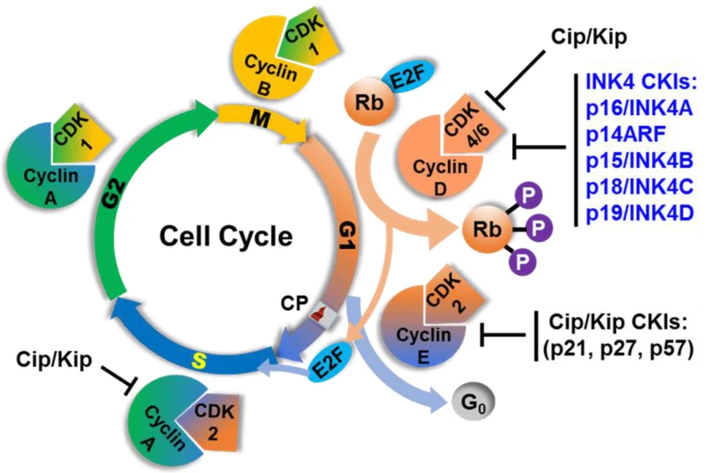
Source: Lutful Kabir, F.M.; Alvarez, C.E.; Bird, R.C. Canine Mammary Carcinomas: A Comparative Analysis of Altered Gene Expression. Vet. Sci. 2016, 3, 1. https://doi.org/10.3390/vetsci3010001
Distinct cyclin–CDK combinations become active at different points in the cell cycle, ensuring orderly progression.
G1 Cyclin–CDK Complexes
Cyclin D associates with CDK4 and CDK6 during early G1 phase.
These complexes:
- Promote progression through G1
- Prepare the cell for DNA replication
- Integrate growth-related signals
Their activity helps the cell commit to entering the replication phase.
G1/S Transition Complexes
Cyclin E binds to CDK2 at the transition from G1 to S phase.
This complex:
- Drives the cell past the restriction point
- Activates proteins required for DNA replication
- Ensures commitment to genome duplication
Once activated, the cell is generally committed to completing the cycle.
S Phase Complexes
Cyclin A partners with CDK2 during S phase.
This complex:
- Regulates DNA replication machinery
- Prevents re-initiation of replication
- Maintains replication fidelity
By controlling replication origin activity, cyclin A–CDK2 ensures that each segment of DNA is replicated exactly once.
Mitotic CDKs
Cyclin B associates with CDK1 to initiate mitosis.
This complex:
- Promotes chromosome condensation
- Facilitates nuclear envelope breakdown
- Activates mitotic spindle formation
Cyclin B–CDK1 acts as the master trigger for entry into mitosis.
Proteolysis and Irreversibility of the Cell Cycle
A key feature of cell cycle progression is directionality. Once a cell moves forward, it does not revert to a previous stage. This irreversibility is largely controlled by regulated protein degradation.
Why Cyclins Must Be Destroyed
If cyclins remained stable:
- CDKs would stay permanently active
- The cell cycle would lose temporal order
- Re-replication or uncontrolled progression could occur
Therefore, cyclins are targeted for destruction once their function is complete.
The Ubiquitin–Proteasome System
Cyclins are marked for degradation by the attachment of ubiquitin molecules. Polyubiquitinated cyclins are recognized and degraded by the proteasome.
This process ensures:
- Rapid inactivation of CDKs
- Clean transitions between phases
- Prevention of backward progression
APC/C Complex
The Anaphase-Promoting Complex/Cyclosome (APC/C) is an E3 ubiquitin ligase that targets specific proteins for degradation during mitosis.
APC/C:
- Promotes separation of sister chromatids
- Triggers degradation of cyclin B
- Drives exit from mitosis
Destruction of cyclin B inactivates CDK1, allowing the cell to complete division.
SCF Complex
The SCF (Skp1–Cullin–F-box) complex is another E3 ubiquitin ligase.
It:
- Regulates G1/S transitions
- Targets specific inhibitors for degradation
- Fine-tunes cyclin–CDK activation
Together, APC/C and SCF ensure that the cell cycle moves forward in a unidirectional manner.
CDKs and Cell Cycle Checkpoints
Cell cycle checkpoints act as surveillance systems that monitor:
- DNA integrity
- Replication completion
- Chromosome alignment
Rather than creating entirely separate pathways, checkpoints often function by modulating CDK activity.
They can:
- Increase CKI levels
- Enhance inhibitory phosphorylation
- Prevent activating phosphorylation
- Delay cyclin synthesis
Through these mechanisms, checkpoints temporarily restrain CDK activity until conditions are appropriate for progression.
This integration ensures fidelity without disrupting overall cycle architecture.
Coordination Between Cell Growth and CDK Activity
Cell division must be coordinated with cell growth. A cell that divides before reaching adequate size risks producing smaller and less viable daughter cells.
Cyclin–CDK activation is influenced by:
- Nutrient availability
- Energy status
- Growth factor signals
- Cellular biosynthetic capacity
Many models suggest a threshold mechanism, where CDK activation requires sufficient accumulation of cyclins. Only when this threshold is reached does the cell commit to progression.
This integration ensures that division reflects the cell’s physiological readiness.
The Integrated Regulatory Network
Cyclins and CDKs do not function in isolation. Their activity emerges from an interconnected network involving:
- Controlled synthesis
- Timed degradation
- Activating phosphorylation
- Inhibitory phosphorylation
- Binding of inhibitors
- Feedback loops
Positive feedback can amplify CDK activation, creating switch-like transitions.
Negative feedback ensures timely inactivation.
This network design provides:
- Precision
- Robustness
- Adaptability
Such regulation allows cells to divide reliably across diverse physiological conditions.
Conclusion
Cyclins and cyclin-dependent kinases form the central regulatory machinery that governs cell division. CDKs provide catalytic power, while cyclins confer timing and specificity. Their activity is shaped by phosphorylation, inhibition, and carefully orchestrated protein degradation.
Through the coordinated action of:
- Phase-specific cyclin synthesis
- Regulated CDK activation
- Targeted proteolysis
- Checkpoint modulation
cells achieve orderly, irreversible, and accurate progression through the cell cycle.
References
Textbooks
- Alberts, B., Johnson, A., Lewis, J., Morgan, D., Raff, M., Roberts, K., & Walter, P. (2022). Molecular Biology of the Cell (7th ed.). Garland Science.
- Lodish, H., Berk, A., Kaiser, C. A., Krieger, M., Bretscher, A., Ploegh, H., Amon, A., & Scott, M. P. (2021). Molecular Cell Biology (9th ed.). W. H. Freeman.
- Cooper, G. M., & Hausman, R. E. (2019). The Cell: A Molecular Approach (8th ed.). Sinauer Associates.
- Morgan, D. O. (2007). The Cell Cycle: Principles of Control. New Science Press.
Educational Resources
- National Center for Biotechnology Information (NCBI) Bookshelf – The Eukaryotic Cell Cycle and Its Control
https://www.ncbi.nlm.nih.gov/books/NBK9876/ - Pellarin, I., Dall’Acqua, A., Favero, A. et al. Cyclin-dependent protein kinases and cell cycle regulation in biology and disease. Sig Transduct Target Ther 10, 11 (2025). https://doi.org/10.1038/s41392-024-02080-z
- Khan Academy – Regulation of the Cell Cycle
https://www.khanacademy.org/science/ap-biology/cell-communication-and-cell-cycle/regulation-of-cell-cycle/a/regulation-of-cell-cycle - Malumbres, M. Cyclin-dependent kinases. Genome Biol 15, 122 (2014). https://doi.org/10.1186/gb4184
Cell cycle regulators are proteins that control progression through G1, S, G2, and M phases. They ensure proper DNA replication and cell division. The main regulators include cyclins, cyclin-dependent kinases (CDKs), and CDK inhibitors.
Cyclins are regulatory proteins whose levels fluctuate during the cell cycle. They bind to and activate CDKs, allowing the cell to progress through specific phases of division.
Cyclin-dependent kinases (CDKs) are enzymes that drive cell cycle transitions. They become active only when bound to a cyclin and then phosphorylate target proteins required for DNA synthesis and mitosis.
Cyclins are produced and degraded at specific stages. Cyclin D acts in G1, Cyclin E at the G1/S transition, Cyclin A during S phase, and Cyclin B controls entry into mitosis.
The four main cyclins are Cyclin D, Cyclin E, Cyclin A, and Cyclin B. Each regulates a distinct phase of the cell cycle by activating specific CDKs.

 regulate the cell cycle, control division timing, and ensure orderly progression in normal cell biology.)