

DNA methylation is one of the most important epigenetic mechanisms involved in the regulation of gene activity. It consists mainly of the addition of a methyl group to cytosine residues, particularly at CpG sites, and can influence whether specific genes are expressed or silenced.
Abnormal DNA methylation is frequently observed in cancer and other diseases. For example, hypermethylation of gene promoter regions can suppress the expression of tumor-suppressor genes and contribute to abnormal cell growth.
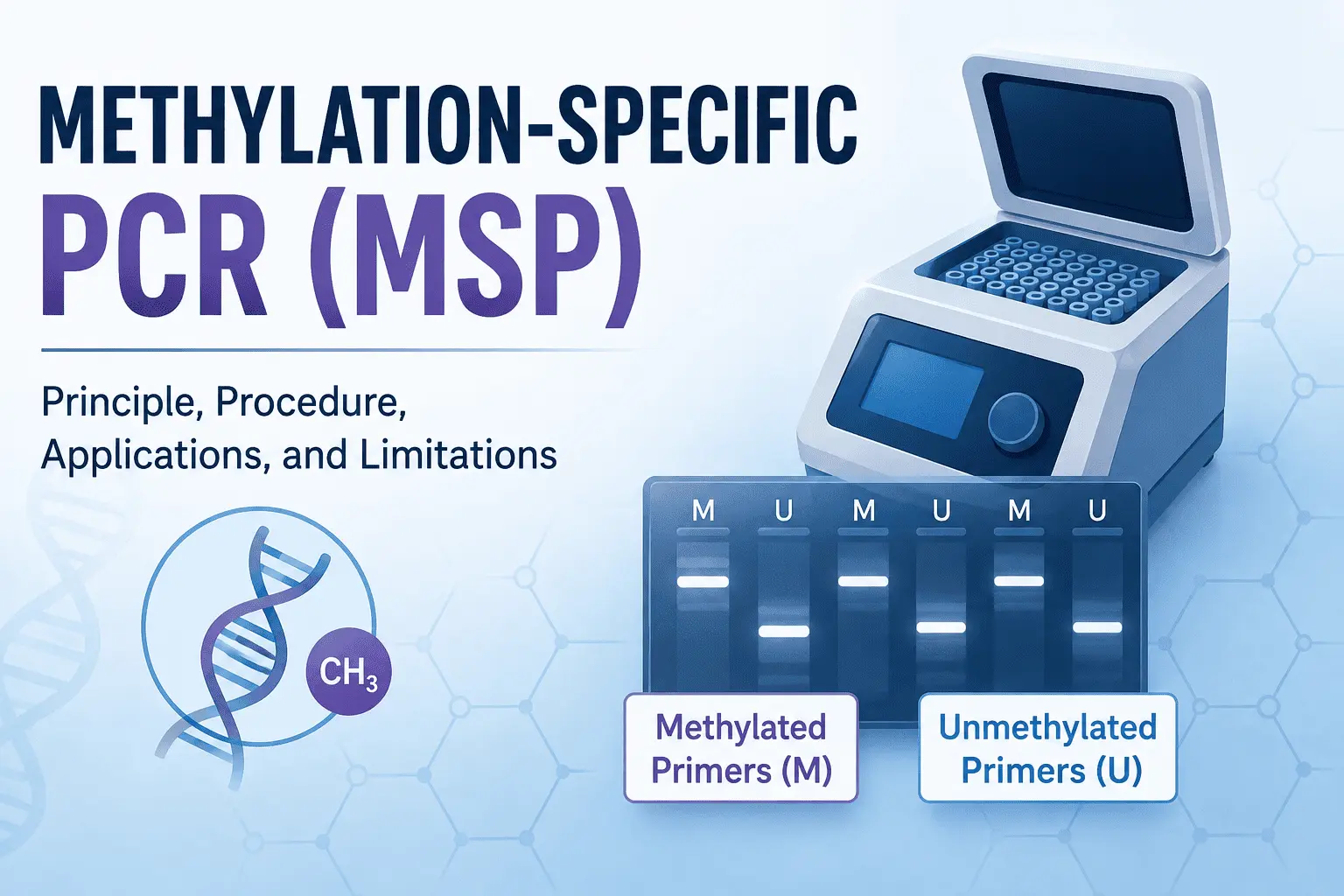
Methylation-Specific PCR (MSP) is a targeted PCR-based technique developed to detect DNA methylation at specific genomic regions. The method combines sodium bisulfite treatment with specially designed primers that distinguish methylated DNA from unmethylated DNA.
MSP is relatively simple, sensitive, and inexpensive, making it useful in molecular biology, epigenetics, cancer research, and biomarker studies.
This article explains the principle of MSP, its workflow, applications, advantages, limitations, and the factors that can affect its results.
What Is Methylation-Specific PCR (MSP)?
Methylation-Specific PCR, commonly abbreviated as MSP, is a PCR technique used to determine whether specific DNA sequences are methylated.
The method relies on a chemical difference between methylated and unmethylated cytosines after bisulfite conversion. During this treatment, unmethylated cytosines are converted into uracils, whereas methylated cytosines are protected from conversion and remain as cytosines.
After PCR amplification, the converted uracils are read as thymine. As a result, bisulfite treatment creates sequence differences between methylated and unmethylated DNA.
These differences allow researchers to design two sets of primers:
- Methylation-specific primers (M primers) recognize the methylated version of the bisulfite-converted DNA.
- Unmethylation-specific primers (U primers) recognize the unmethylated version.
PCR is then performed separately with the two primer pairs. The amplification pattern provides information about the methylation status of the target region.
DNA methylation and CpG sites
DNA methylation in mammals occurs predominantly at cytosine residues followed by guanine, known as CpG sites.
Clusters of CpG sites, called CpG islands, are frequently found near gene promoters. In many biological contexts, methylation of promoter-associated CpG sites is associated with reduced transcription.
This relationship is particularly important in cancer research. Tumor cells can acquire abnormal methylation patterns that silence genes involved in cell-cycle regulation, DNA repair, apoptosis, and other cellular processes.
The basic principle of MSP
The principle can be summarized as follows:
Genomic DNA → bisulfite conversion → methylated/unmethylated sequence differences → methylation-specific PCR → detection of amplification
If the methylation-specific reaction produces a PCR product, methylated DNA is present in the analyzed sample. If the unmethylation-specific reaction produces a product, unmethylated DNA is present.
When both reactions produce products, the sample may contain both methylated and unmethylated copies of the target sequence.
MSP is therefore a targeted methylation assay. It does not examine methylation throughout the entire genome but instead focuses on specific genomic regions selected by the researcher.
How Does Methylation-Specific PCR Work?
The MSP procedure consists of several interconnected steps, beginning with DNA extraction and ending with interpretation of the PCR products.
1. DNA extraction
The first step is to isolate genomic DNA from the biological sample.
Depending on the study, DNA can be extracted from sources such as:
- Tumor tissue
- Normal tissue
- Blood
- Cultured cells
- Body fluids
- Biopsy specimens
DNA quality is particularly important because bisulfite treatment can cause DNA degradation. Starting with highly degraded or contaminated DNA can therefore reduce the efficiency of the subsequent MSP reaction.
The extracted DNA should generally be evaluated for concentration and purity before bisulfite conversion.
2. Bisulfite conversion
Bisulfite conversion is the critical chemical step that makes methylation-specific detection possible.
During treatment with sodium bisulfite under appropriate conditions, unmethylated cytosines are converted to uracils, while 5-methylcytosines remain largely unchanged.
The resulting sequence differences can subsequently be detected by PCR.
For example, consider a simplified DNA sequence containing a CpG site:
Before conversion:
...CG...
If the cytosine is unmethylated, bisulfite treatment converts it:
Unmethylated DNA:
...TG...
If the cytosine is methylated, it remains:
Methylated DNA:
...CG...
PCR primers can therefore be designed to discriminate between these two sequences.
The efficiency of bisulfite conversion is extremely important. If unmethylated cytosines fail to convert, they may incorrectly appear to be methylated, producing a false-positive methylation result.
At the same time, bisulfite treatment can fragment or damage DNA, meaning that the conversion procedure needs to be optimized for the sample type and downstream assay.
3. MSP primer design
Following bisulfite conversion, primers are designed specifically for the methylated and unmethylated versions of the target sequence.
Usually, two primer pairs are used:
Methylated-specific primer pair
This primer pair is designed to amplify the target sequence when the relevant CpG positions remain as cytosines after bisulfite treatment.
Unmethylated-specific primer pair
This primer pair is designed to recognize the sequence in which the corresponding unmethylated cytosines have been converted.
Primer design is one of the most important determinants of MSP specificity.
Primers should be designed around informative CpG sites so that the methylated and unmethylated sequences can be distinguished. Other factors, including primer length, melting temperature, GC content, potential secondary structures, and amplicon size, must also be considered.
The selected region should be sufficiently short to accommodate the fragmented DNA that can result from bisulfite treatment.
4. PCR amplification
After bisulfite conversion and primer preparation, the modified DNA is amplified by PCR.
The methylated and unmethylated reactions can be performed separately using their respective primer pairs.
Like conventional PCR, MSP amplification involves repeated cycles of:
- Denaturation – separation of the DNA strands.
- Annealing – binding of the primers to their complementary sequences.
- Extension – synthesis of new DNA by DNA polymerase.
The exact cycling conditions depend on the primers, polymerase, template, and experimental system and should be optimized experimentally.
Appropriate controls are also essential.
A no-template control (NTC) can help detect contamination, while methylated and unmethylated control DNA can help evaluate whether the respective primer systems are working correctly.
5. Detection of PCR products
In conventional MSP, PCR products are commonly analyzed using agarose gel electrophoresis.
The amplified DNA fragments are separated according to size and visualized as bands.
A band in the methylation-specific reaction indicates that the methylated sequence was amplified. Similarly, a band in the unmethylation-specific reaction indicates amplification of the unmethylated sequence.
The expected PCR product size should be confirmed by comparison with an appropriate DNA size marker.
6. Interpretation of MSP results
The interpretation of MSP depends on the amplification pattern obtained from the M and U reactions.
A simplified interpretation is:
| Methylation-specific PCR | Unmethylation-specific PCR | Interpretation |
|---|---|---|
| Positive | Negative | Methylated DNA detected |
| Negative | Positive | Unmethylated DNA detected |
| Positive | Positive | Both methylated and unmethylated DNA detected |
| Negative | Negative | No detectable amplification or technically inconclusive |
For example, if a tumor sample produces a strong band with the methylated primers but no band with the unmethylated primers, the target region is considered methylated according to the assay.
However, MSP results should not automatically be interpreted as an exact measurement of methylation percentage.
A positive MSP result indicates that DNA molecules compatible with the methylated primer sequence are present. It does not necessarily mean that every copy of the target region is methylated.
This distinction becomes particularly important in heterogeneous biological samples, such as tumors containing mixtures of cancer cells, stromal cells, and immune cells.
Methylation-Specific PCR Applications in Research and Medicine
MSP has been widely used to investigate DNA methylation in different biological and clinical contexts.
Cancer research
Cancer research is one of the most important applications of MSP.
Tumor cells can undergo extensive epigenetic alterations, including abnormal methylation of gene promoters. When regulatory regions of tumor-suppressor genes become hypermethylated, transcription can be reduced or silenced.
MSP can be used to determine whether a particular gene promoter is methylated in tumor samples.
Researchers can compare methylation patterns between:
- Tumor and normal tissues
- Different cancer subtypes
- Primary and metastatic tumors
- Treatment-sensitive and treatment-resistant samples
- Early- and advanced-stage disease
This makes MSP useful for investigating relationships between DNA methylation and cancer development.
Gene regulation and epigenetics
MSP can also help researchers investigate the relationship between DNA methylation and gene expression.
For example, a researcher may want to determine whether reduced expression of a particular gene is associated with promoter methylation.
In such a study, MSP can be combined with gene-expression techniques such as RT-PCR or quantitative PCR.
If a gene shows reduced expression and increased promoter methylation, the results may support a relationship between epigenetic modification and transcriptional regulation. However, MSP alone cannot establish that methylation directly caused the change in gene expression.
Biomarker discovery
DNA methylation patterns can be investigated as potential biomarkers for different diseases.
In cancer, researchers may examine whether methylation of a particular genomic region occurs more frequently in patients than in healthy controls.
Potential applications include:
- Diagnostic biomarker research
- Prognostic biomarker research
- Disease classification
- Treatment-response studies
- Disease monitoring
MSP is attractive for targeted biomarker studies because once a methylated region has been identified, the assay can be applied to many samples relatively efficiently.
Analysis of biological samples
Another important advantage of MSP is its ability to investigate methylation in different sample types.
Depending on the research question and DNA quality, studies can analyze methylation in tissue specimens, blood-derived DNA, cultured cells, and other biological materials.
This has contributed to research into circulating DNA and minimally invasive approaches to disease biomarker detection.
Validation of methylation findings
MSP can also be used to validate candidate methylation changes discovered using other approaches.
For example, a large-scale methylation analysis may identify a genomic region that appears to be differentially methylated between two groups.
Researchers can subsequently develop a targeted MSP assay to investigate that region in an independent set of samples.
This targeted approach can be particularly useful when a study involves a relatively large number of samples but only a limited number of genomic regions.
Advantages, Limitations, and Alternatives to MSP
MSP remains a useful technique because it combines PCR with targeted methylation detection. However, it also has important limitations that should be considered when selecting a methylation-analysis method.
Advantages of MSP
One of the main advantages of MSP is its simplicity.
The method uses equipment commonly available in molecular biology laboratories, including standard PCR instruments and gel electrophoresis systems.
Other advantages include:
- High sensitivity for targeted methylation detection
- Relatively low cost
- Straightforward experimental workflow
- Ability to analyze specific genomic regions
- Compatibility with many biological sample types
- Suitability for relatively large numbers of samples
- Useful application in cancer and epigenetic research
Because MSP focuses on a predefined genomic region, it can also be more practical than genome-wide approaches when the research question concerns a small number of candidate genes.
Limitations of MSP
Despite these advantages, conventional MSP has several limitations.
Limited quantitative information
Standard MSP is primarily a qualitative or semi-quantitative technique.
A positive methylation-specific PCR reaction indicates the presence of methylated DNA but does not directly provide an accurate percentage of methylation.
When precise methylation levels are required, quantitative approaches may be more appropriate.
Dependence on bisulfite conversion
The accuracy of MSP depends heavily on the efficiency of bisulfite conversion.
Incomplete conversion can cause unmethylated cytosines to remain unchanged, potentially causing them to be interpreted as methylated.
Therefore, conversion efficiency should be carefully considered when designing and interpreting an MSP experiment.
Primer specificity
MSP requires primers that effectively distinguish methylated from unmethylated sequences.
Poorly designed primers can produce non-specific amplification or preferentially amplify one DNA population.
Primer design and PCR optimization are therefore critical.
Limited genomic coverage
MSP is a targeted technique. It generally analyzes only the region selected during primer design.
It cannot provide a comprehensive picture of methylation throughout the genome.
DNA degradation
Bisulfite treatment can damage and fragment DNA. This can be particularly problematic when working with limited or poor-quality samples.
Cell heterogeneity
Clinical samples may contain several different cell populations.
For example, a tumor specimen can contain cancer cells alongside normal cells, immune cells, fibroblasts, and vascular cells.
A mixed MSP signal may therefore reflect the composition of the sample rather than uniform methylation within every cell.
Common causes of unsuccessful MSP
Several technical problems can affect MSP results.
Poor DNA quality: Degraded DNA may produce weak or inconsistent amplification.
Incomplete bisulfite conversion: Unconverted cytosines can lead to incorrect methylation calls.
Poor primer design: Primers that do not discriminate efficiently between methylated and unmethylated sequences can generate misleading results.
Non-specific amplification: Incorrect PCR conditions may result in unexpected bands.
PCR inhibition: Contaminants carried over during DNA extraction can interfere with amplification.
Contamination: Because PCR is highly sensitive, contamination can generate false-positive results.
Insufficient template: Very low DNA concentrations can produce weak or absent amplification.
Including appropriate controls and optimizing both conversion and PCR conditions can help identify these problems.
Alternatives to methylation-specific PCR
MSP is not the only method available for DNA methylation analysis.
Researchers may select other techniques depending on whether they need qualitative, quantitative, locus-specific, or genome-wide information.
Quantitative MSP (qMSP) extends the MSP concept by using real-time PCR to quantify methylated DNA more precisely.
Bisulfite sequencing combines bisulfite conversion with DNA sequencing and can provide methylation information at individual cytosine positions across a targeted region.
Pyrosequencing can provide quantitative methylation measurements at selected CpG sites and is useful when precise methylation percentages are required.
Methylation-sensitive restriction enzyme methods use enzymes whose activity depends on the methylation status of specific recognition sites.
DNA methylation arrays allow researchers to investigate methylation at large numbers of predefined genomic sites.
Next-generation sequencing-based approaches can provide much broader methylation profiles and are useful for large-scale or genome-wide studies.
The choice between these approaches depends on the research objective, required resolution, number of samples, available equipment, and budget.
Conclusion
Methylation-Specific PCR (MSP) is a targeted PCR-based method used to detect DNA methylation at specific genomic regions. Its fundamental principle relies on bisulfite conversion, which creates sequence differences between methylated and unmethylated DNA that can subsequently be distinguished using methylation-specific primers.
The workflow generally involves DNA extraction, bisulfite conversion, primer design, PCR amplification, detection of PCR products, and interpretation of methylation-specific and unmethylation-specific amplification.
MSP has important applications in cancer research, epigenetics, gene regulation, and biomarker discovery. Its relatively simple workflow, sensitivity, and low cost make it particularly useful for targeted studies involving many samples.
However, MSP also has limitations. Conventional MSP does not provide precise methylation percentages, depends strongly on efficient bisulfite conversion and primer specificity, and provides information only about selected genomic regions.
When more detailed information is required, techniques such as qMSP, bisulfite sequencing, pyrosequencing, methylation arrays, or sequencing-based approaches may be more appropriate.
Overall, MSP remains a valuable technique for researchers who need a practical method to investigate methylation at specific DNA regions, particularly when studying candidate genes or validating methylation biomarkers.


 works, including bisulfite conversion, primer design, PCR steps, applications, advantages, limitations, and results.)