

Key Takeaways
- MYC is a major oncogene that regulates cell growth, proliferation, metabolism, and protein synthesis.
- The MYC gene on chromosome 8q24 encodes the c-Myc transcription factor, which mainly functions by forming a complex with MAX.
- MYC alterations in cancer include gene amplification, chromosomal translocations, enhancer activation, and abnormal signaling—not just mutations.
- MYC amplification can increase MYC expression and activity and is observed in several solid and hematological cancers.
- The MYC family includes MYC (c-Myc), MYCN (N-Myc), and MYCL (L-Myc), each associated with different cancer types.
- Abnormally high MYC activity promotes cell proliferation, metabolic reprogramming, ribosome biogenesis, and genomic instability.
- MYC can contribute to tumor progression, immune evasion, metastasis, and resistance to cancer treatments.
- MYC activity signatures may provide useful information for cancer prognosis and treatment research, although MYC is not yet a universal standalone clinical biomarker.
- MYC has historically been considered difficult to target, but new strategies are emerging, including direct MYC inhibition, protein degradation, and MYC RNA targeting.
- Emerging MYC-targeted therapies such as OMO-103 and MYC-degrading or RNA-targeting approaches are being investigated, offering potential new strategies for MYC-dependent cancers.
In this article, we explore the MYC oncogene, its structure and functions, cancer-associated alterations, role in tumor progression, and the latest advances in MYC-targeted therapies.
1. MYC Gene and Protein
What Is the MYC Gene?
MYC, officially called MYC proto-oncogene, bHLH transcription factor, is located on chromosome 8q24.21. It belongs to the proto-oncogene class, meaning that its normal activity contributes to physiological processes such as cell growth and proliferation but can promote cancer when abnormally activated.
The protein encoded by MYC is known as c-Myc. MYC expression is normally induced when cells receive growth-promoting signals and decreases when those signals disappear.
MYC is therefore a tightly regulated component of normal cellular physiology. Cancer cells can disrupt this regulation, resulting in persistent MYC activity.
The c-Myc Protein
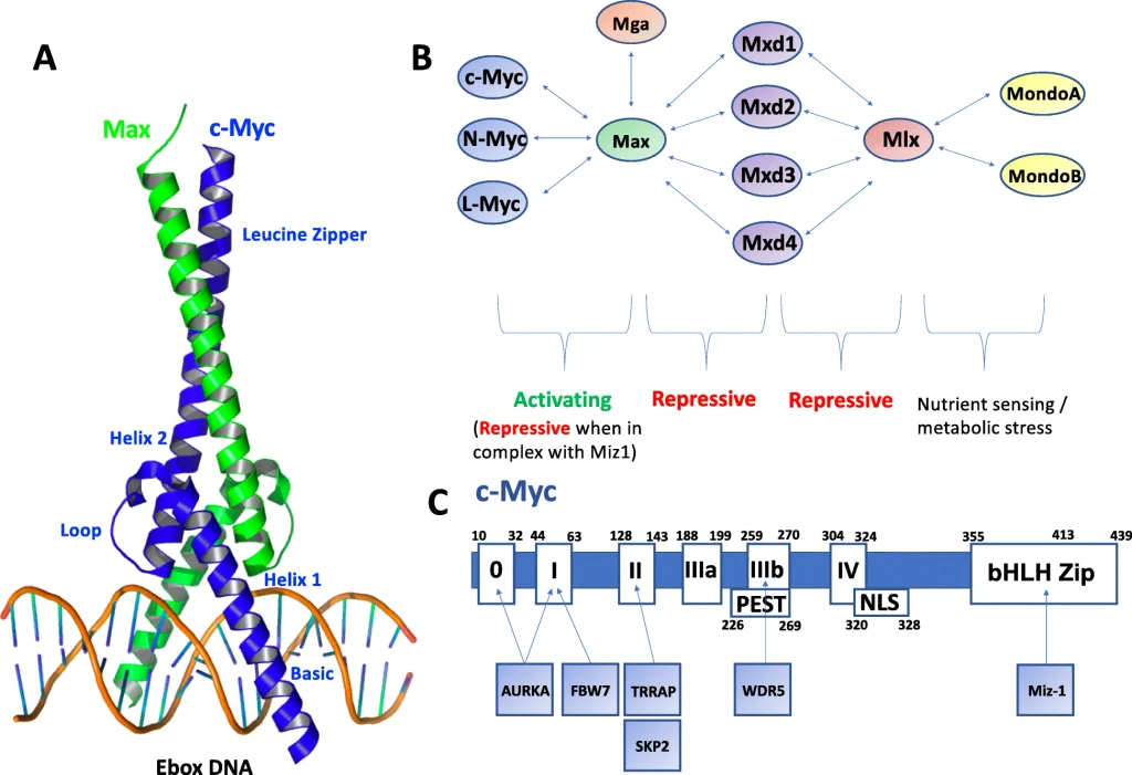
Madden, S.K., de Araujo, A.D., Gerhardt, M. et al. Taking the Myc out of cancer: toward therapeutic strategies to directly inhibit c-Myc. Mol Cancer 20, 3 (2021). https://doi.org/10.1186/s12943-020-01291-6
c-Myc is a basic helix-loop-helix leucine zipper (bHLH-LZ) transcription factor. One of its most important partners is MAX.
The MYC–MAX complex binds specific DNA sequences known as E-boxes and regulates the transcription of numerous genes.
Through this mechanism, MYC influences:
- Cell-cycle progression
- DNA replication
- Ribosome biogenesis
- Protein synthesis
- Glucose and glutamine metabolism
- Nucleotide synthesis
- Mitochondrial function
- Cell growth
- Apoptosis
MYC therefore acts as a broad transcriptional regulator rather than controlling a single biological pathway.
The MYC Family
The human MYC family contains three major members:
MYC encodes c-Myc and is involved in a broad range of cancers.
MYCN encodes N-Myc and is particularly important in neuroblastoma, where MYCN amplification is associated with aggressive disease.
MYCL encodes L-Myc and has been associated with cancers including small-cell lung cancer.
Although MYC, MYCN, and MYCL share important structural features, their tissue distributions and cancer associations differ.
Normal Functions of MYC
Under physiological conditions, MYC helps coordinate cellular growth with the production of energy and macromolecules required for proliferation.
It stimulates cell-cycle progression, ribosome production, protein synthesis, and metabolic pathways.
This explains an important feature of MYC biology: the same programs that support normal growth can become powerful drivers of cancer when MYC remains continuously activated.
2. MYC Alterations in Cancer
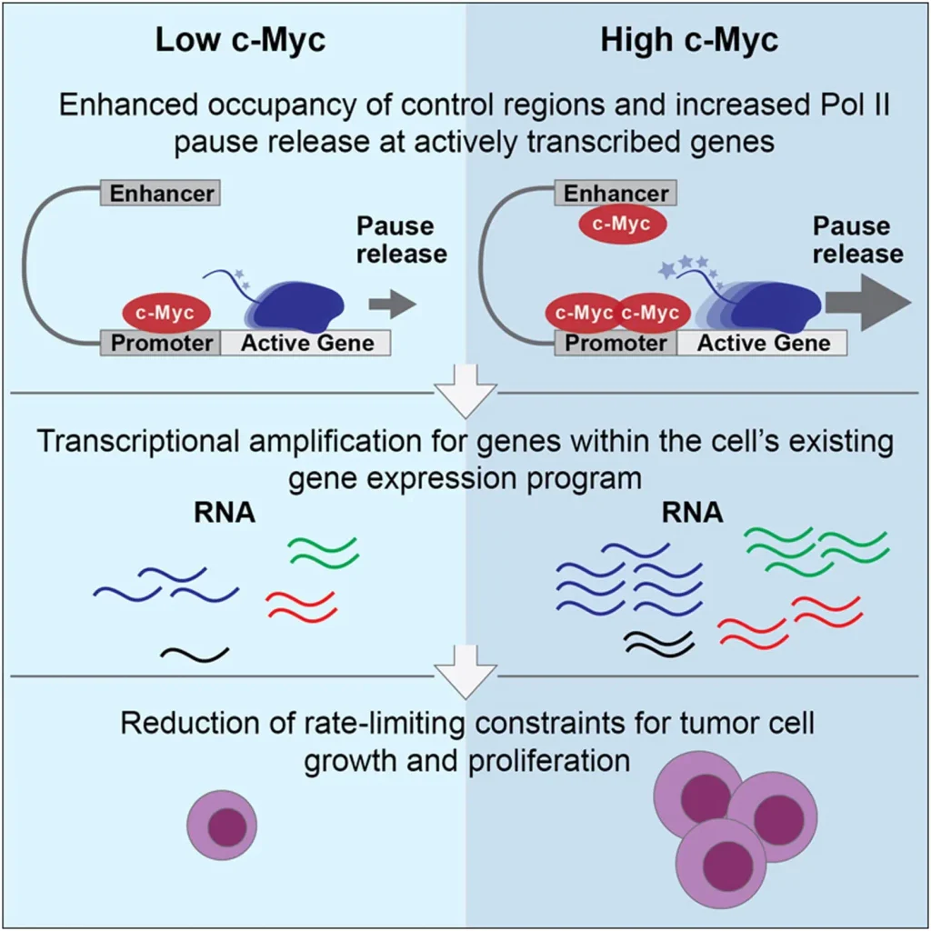
Lin CY, Lovén J, Rahl PB, Paranal RM, Burge CB, Bradner JE, Lee TI, Young RA. Transcriptional amplification in tumor cells with elevated c-Myc. Cell. 2012 Sep 28;151(1):56-67. doi: 10.1016/j.cell.2012.08.026.
MYC can become oncogenic through several mechanisms. Importantly, MYC activation does not necessarily require a mutation in the MYC gene.
MYC Amplification
MYC amplification occurs when cancer cells acquire additional copies of the MYC-containing genomic region.
Increased MYC copy number can lead to increased MYC expression and transcriptional activity.
MYC amplification has been reported in several cancers, including subsets of:
- Breast cancer
- Colorectal cancer
- Ovarian cancer
- Pancreatic cancer
- Lung cancer
- Brain tumors
- Osteosarcoma
The extent and structure of amplification can vary between tumors.
Importantly, amplification should not automatically be interpreted as equivalent to MYC dependency. Recent large-scale genomic research involving more than 68,000 tumor-normal samples found that MYC amplification is much more common than MYCN or MYCL amplification in many solid tumors, while also showing that functional MYC dependency depends on additional characteristics such as tumor lineage and coexisting genomic alterations.
This distinction is important for future MYC-targeted therapies.
MYC Mutations
Unlike genes such as KRAS, MYC is not usually activated through recurrent classical activating mutations.
Instead, MYC is more commonly deregulated through amplification, rearrangement, enhancer activation, abnormal transcription, altered protein stability, or activation of upstream signaling pathways.
Nevertheless, somatic MYC mutations can occur and may influence protein stability, transcriptional activity, or interactions with regulatory proteins. Such mutations have attracted particular interest in certain hematological malignancies.
Therefore, MYC cancer biology should not be evaluated by mutation status alone.
MYC Translocations
One of the classic examples of MYC activation occurs in Burkitt lymphoma.
The characteristic rearrangement is:
t(8;14)
This places MYC near the immunoglobulin heavy-chain locus, exposing MYC to strong regulatory elements that drive abnormal expression.
Other MYC translocations can involve immunoglobulin light-chain loci.
Persistent MYC expression contributes to the extremely rapid proliferation characteristic of Burkitt lymphoma.
Enhancer Activation and Upstream Signaling
MYC can also be activated without amplification or mutation.
Cancer-associated changes in enhancers and chromatin organization can increase MYC transcription. Some tumors can develop highly active regulatory regions known as super-enhancers that promote strong oncogene expression.
MYC is also an important downstream target of several oncogenic pathways:
WNT/β-catenin → MYC
RAS/MAPK → MYC
PI3K/AKT/mTOR → MYC
NOTCH → MYC
JAK/STAT → MYC
Consequently, MYC can function as a convergence point for multiple cancer-driving signals.
3. How MYC Promotes Cancer Development and Progression
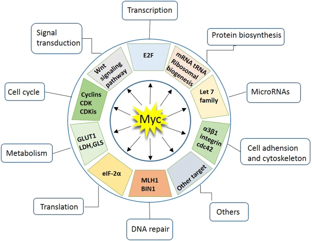
Chen, H., Liu, H. & Qing, G. Targeting oncogenic Myc as a strategy for cancer treatment. Sig Transduct Target Ther 3, 5 (2018). https://doi.org/10.1038/s41392-018-0008-7
Persistent MYC activation affects many biological processes that support tumor growth.
MYC and Cell Proliferation
MYC stimulates the expression of genes involved in cell-cycle progression and DNA replication.
In cancer cells, continuous MYC activity can therefore promote repeated cell division and contribute to uncontrolled tumor growth.
MYC and Cancer Metabolism
Rapidly growing cancer cells require large quantities of energy and biosynthetic substrates.
MYC promotes metabolic reprogramming by increasing processes such as:
- Glucose uptake
- Glycolysis
- Glutamine utilization
- Nucleotide synthesis
- Amino-acid metabolism
- Protein synthesis
This allows MYC-driven cells to generate the energy and building blocks required for rapid proliferation.
However, this metabolic reprogramming can also create vulnerabilities. MYC-dependent tumors may become unusually reliant on specific metabolic pathways, providing potential therapeutic targets.
MYC and Ribosome Biogenesis
MYC stimulates ribosome biogenesis, allowing cancer cells to increase their capacity for protein synthesis.
The relationship can be summarized as:
MYC activation → ribosome production → protein synthesis → cellular growth
Because MYC-driven cells often have exceptionally high protein-production requirements, ribosome biogenesis and translational machinery are being investigated as potential therapeutic vulnerabilities.
MYC, Replication Stress, and Genomic Instability
Excessive MYC activity can accelerate DNA replication and increase replication stress.
This can result in:
- DNA damage
- Replication abnormalities
- Genomic instability
MYC-driven cancer cells may consequently become dependent on DNA damage-response pathways that help them survive replication stress.
This creates opportunities for synthetic-lethal treatment strategies.
MYC and Apoptosis
MYC has a complex relationship with apoptosis.
Although it promotes cell proliferation, excessive MYC activity can also trigger apoptotic pathways when appropriate survival signals are absent.
Cancer cells can overcome this barrier through additional alterations that increase survival signaling or disrupt tumor-suppressive pathways.
Thus, MYC frequently cooperates with other oncogenic or tumor-suppressive alterations during tumor development.
MYC and the Tumor Microenvironment
MYC activity is increasingly recognized as an important regulator of the tumor microenvironment.
It can influence cytokine signaling, immune-cell recruitment, macrophage behavior, and other processes that affect the interaction between cancer cells and the immune system.
This has created interest in combining MYC-targeted strategies with immunotherapy.
MYC and Treatment Resistance
MYC can contribute to treatment resistance through several mechanisms, including:
- Increased proliferation
- Metabolic adaptation
- Resistance to apoptosis
- Replication-stress responses
- Cancer-cell plasticity
- Immune evasion
Consequently, targeting MYC or MYC-dependent pathways could potentially improve responses to existing cancer treatments.
4. MYC in Cancer Diagnosis, Prognosis, and Therapeutic Research
MYC has attracted considerable interest as both a biomarker and therapeutic target.
Detecting MYC Alterations
MYC can be investigated at several molecular levels.
DNA analysis can identify amplification, copy-number alterations, and chromosomal rearrangements.
RNA analysis can measure MYC transcription using techniques such as RT-qPCR or RNA sequencing.
Protein analysis can evaluate c-Myc expression using methods such as immunohistochemistry or immunoblotting.
Researchers are also developing MYC transcriptional signatures that may provide information about pathway activity rather than simply MYC expression.
This distinction is increasingly important because MYC amplification does not always indicate that a tumor is functionally dependent on MYC.
MYC and Cancer Prognosis
High MYC activity has been associated with aggressive disease in several cancers.
Depending on tumor type, MYC deregulation may correlate with:
- Increased proliferation
- Tumor progression
- Metastasis
- Treatment resistance
- Poor clinical outcomes
However, the prognostic significance of MYC varies between cancers and should be interpreted in the context of other molecular and clinical characteristics.
5. Emerging MYC-Targeted Therapies and Latest Research
For decades, MYC was described as an “undruggable” oncogene. Its intrinsically disordered regions and reliance on protein–protein interactions made conventional drug development difficult.
Recent research is changing this view.
Direct MYC Inhibition
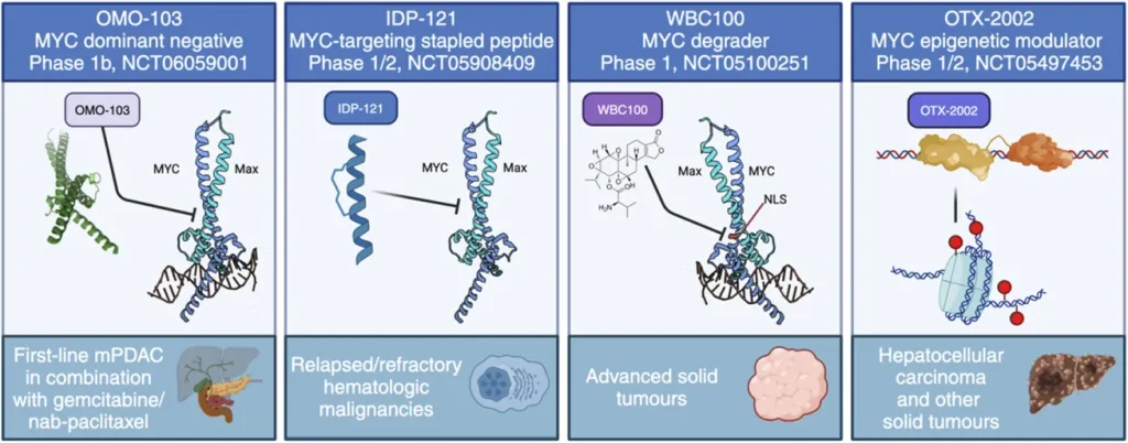
Zacarías-Fluck MF, Soucek L and Whitfield JR (2024) MYC: there is more to it than cancer. Front. Cell Dev. Biol. 12:1342872. doi: 10.3389/fcell.2024.1342872
One approach is to interfere with the interaction between MYC and MAX.
Blocking the MYC–MAX complex can reduce MYC-dependent transcription and potentially suppress tumor growth.
One of the most advanced approaches is Omomyc, an engineered MYC-derived protein designed to interfere with MYC function.
Its clinical formulation, OMO-103, has entered human clinical testing. Early studies have demonstrated MYC pathway inhibition and preliminary evidence of antitumor activity, although OMO-103 remains investigational.
MYC Protein Degradation
Instead of simply inhibiting MYC, researchers are developing approaches designed to destroy the MYC protein.
The concept is:
MYC → targeted degradation → MYC protein ↓ → MYC signaling ↓
Strategies under investigation include targeted protein degradation and approaches that destabilize proteins required for MYC stability.
This could potentially produce deeper suppression of MYC signaling than conventional inhibition.
Targeting MYC RNA
One of the most innovative recent directions is targeting MYC mRNA.
In 2023, researchers reported a MYC-targeting RNA degrader known as MYC-RiboTAC, designed to recruit endogenous RNase L and promote degradation of MYC mRNA. More recently, this strategy was investigated in multiple myeloma models, where MYC-RiboTAC showed promising preclinical antitumor activity.
This represents a fundamentally different strategy:
Instead of targeting the difficult-to-drug MYC protein, destroy the RNA that produces it.
Targeting MYC Regulators
Another strategy is to target proteins that control MYC expression or stability.
Researchers are investigating regulators involved in:
- MYC transcription
- MYC translation
- MYC protein stability
- MYC degradation
- MYC-associated chromatin regulation
Examples include BET proteins, transcriptional regulators, kinases, and RNA-binding proteins.
This indirect approach may be useful because some MYC regulators have structures that are more amenable to conventional drug development.
Targeting MYC-Dependent Vulnerabilities
Perhaps the most important long-term strategy is synthetic lethality.
Instead of directly inhibiting MYC, researchers can identify pathways that MYC-driven cancer cells become dependent upon.
Potential vulnerabilities include:
- DNA damage-response pathways
- Replication-stress responses
- Metabolic pathways
- Protein synthesis
- Ribosome biogenesis
- Proteostasis
The concept is:
MYC activation → cellular dependency → therapeutic inhibition → selective cancer-cell death
This strategy may be particularly useful because completely blocking MYC could also affect normal cells that require MYC for physiological growth.
MYC and Combination Therapy
Future MYC-directed treatments will likely involve combinations rather than MYC inhibition alone.
Potential combinations include:
MYC inhibition + chemotherapy
MYC inhibition + targeted therapy
Immunotherapy + MYC inhibition
MYC inhibition + metabolic inhibitors
DNA-damage-response inhibitors + MYC inhibition
The goal is to exploit the biological vulnerabilities created by MYC while reducing the possibility of resistance.
References and Further Reading
- Tong, Y., Lee, Y., Liu, X. et al. Programming inactive RNA-binding small molecules into bioactive degraders. Nature 618, 169–179 (2023). https://doi.org/10.1038/s41586-023-06091-8.
- Maisano D, Wang T, Kulp SB, Favasuli VK, Tong Y, Cumerlato M, Li C, Barello F, Turi M, Rampa P, Parisi RA, Garbicz F, Ladisa F, Giorgetti G, Chakraborty C, Vreux L, Sangiorgio V, Sarotto I, Aktas Samur A, Samur MK, Carrasco RD, Fulciniti M, Cameron MD, Anderson KC, Gulla A, Childs-Disney J, Disney MD, Morelli E, Munshi NC. A first-in-class RNA degrader reduces c-MYC expression and myeloma cell survival in preclinical models. Blood. 2026 Jul 29:blood.2026033241. https://doi.org/10.1182/blood.2026033241.
- Zacarías-Fluck MF, Soucek L and Whitfield JR (2024) MYC: there is more to it than cancer. Front. Cell Dev. Biol. 12:1342872. https://doi.org/10.3389/fcell.2024.1342872
- Chen, H., Liu, H. & Qing, G. Targeting oncogenic Myc as a strategy for cancer treatment. Sig Transduct Target Ther 3, 5 (2018). https://doi.org/10.1038/s41392-018-0008-7.
- Lin CY, Lovén J, Rahl PB, Paranal RM, Burge CB, Bradner JE, Lee TI, Young RA. Transcriptional amplification in tumor cells with elevated c-Myc. Cell. 2012 Sep 28;151(1):56-67. https://doi.org/10.1016/j.cell.2012.08.026.
- Duffy, M.J., Tang, M. & Crown, J. MYC as a Target for Cancer Treatment: from Undruggable to Druggable?. Targ Oncol 20, 791–801 (2025). https://doi.org/10.1007/s11523-025-01169-x.
- Whitfield, J.R., Soucek, L. MYC in cancer: from undruggable target to clinical trials. Nat Rev Drug Discov 24, 445–457 (2025). https://doi.org/10.1038/s41573-025-01143-2.


