

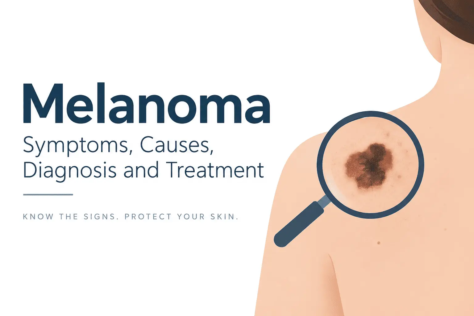
Melanoma is a type of skin cancer that develops from melanocytes, the cells responsible for producing melanin, the pigment that gives skin its color. Although melanoma is less common than some other types of skin cancer, it can be more likely to grow and spread to other parts of the body if it is not detected and treated early.
The good news is that melanoma can often be treated successfully, particularly when it is found at an early stage. Knowing what melanoma looks like, understanding the risk factors, and paying attention to changes in your skin can help with early detection.
What Is Melanoma?
Melanoma is a cancer that begins in melanocytes, which normally produce the pigment melanin. Most melanomas develop in the skin, but melanoma can also occur in other areas where melanocytes are found, including the eyes and, more rarely, mucosal tissues.
Melanoma can develop in a completely new spot or begin in an existing mole. It may appear as a dark or irregularly colored spot, although not all melanomas are black or brown. Some can be pink, red, or have very little pigment.
Unlike a normal mole, a melanoma may gradually change in size, shape, color, or appearance. These changes are important warning signs that should be evaluated by a healthcare professional.
Melanoma is different from other common skin cancers, such as basal cell carcinoma and squamous cell carcinoma. While these cancers are more common, melanoma has a greater tendency to invade deeper tissues and spread to distant organs.
This is why recognizing suspicious skin changes and seeking medical advice early is important.
What Causes Melanoma and Who Is at Risk?
Melanoma develops when melanocytes acquire genetic changes that cause them to grow and divide abnormally. Several factors can contribute to the development of these changes.
Ultraviolet radiation
Exposure to ultraviolet (UV) radiation from sunlight is one of the major environmental risk factors for melanoma. Repeated intense sun exposure and episodes of sunburn, particularly during childhood and adolescence, can increase the risk.
Artificial sources of UV radiation, such as tanning beds, can also contribute to melanoma risk.
However, melanoma does not develop only in areas that receive a lot of sunlight. It can occur on parts of the body that are not regularly exposed to the sun.
Skin type and physical characteristics
Certain physical characteristics are associated with a higher risk of melanoma. These include:
- Fair skin that burns easily
- Light-colored eyes
- Blond or red hair
- A tendency to develop freckles
- Difficulty tanning
Having darker skin does not eliminate the risk. Melanoma can occur in people of all skin tones, and in people with darker skin it may sometimes develop in less obvious locations, such as the palms of the hands, soles of the feet, or beneath the nails.
Moles and previous skin cancer
Having a large number of moles can increase the likelihood of developing melanoma. Certain atypical or unusual-looking moles may also be associated with increased risk.
People who have previously had melanoma or another type of skin cancer may also have a higher risk of developing melanoma in the future.
Family history and genetics
A family history of melanoma can increase risk. In some families, inherited genetic changes can contribute to a higher susceptibility to melanoma.
However, having a family member with melanoma does not mean that a person will necessarily develop the disease.
What Does Melanoma Look Like? Signs and Symptoms
One of the most useful things you can do is become familiar with the appearance of your own skin. A suspicious lesion may look different from your other moles or may change over time.
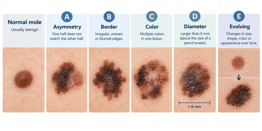
A commonly used method for recognizing possible melanoma is the ABCDE rule.
A — Asymmetry
A mole is considered more concerning when one half does not look similar to the other half.
B — Border
A suspicious lesion may have an irregular, uneven, blurred, or poorly defined border rather than a smooth, regular edge.
C — Color
Melanomas may contain several different colors. A lesion may include shades of brown, black, red, pink, white, or blue.
However, color alone does not determine whether a lesion is melanoma. Some melanomas may have relatively little pigment.
Some melanomas produce little or no pigment and may therefore be harder to recognize; learn more about amelanotic melanoma and how it can appear differently from typical pigmented melanoma.
D — Diameter
Larger lesions can be more concerning, particularly those measuring more than about 6 millimeters. However, melanoma can also be smaller when it is first detected.
Therefore, a small lesion should not automatically be considered harmless.
E — Evolving
One of the most important warning signs is change over time.
A mole or skin lesion that changes in size, shape, color, or appearance should be evaluated. New symptoms such as itching, bleeding, crusting, or persistent irritation can also be reasons to seek medical advice.
The “ugly duckling” sign
Another useful concept is the ugly duckling sign. This refers to a mole that looks noticeably different from the person’s other moles.
For example, if most of your moles have a similar appearance but one is much darker, larger, or more irregular, it deserves closer attention.
Melanoma can also develop in areas that are easy to overlook, including the scalp, between the toes, palms, soles, and underneath fingernails or toenails.
Not every unusual-looking mole is melanoma, and some melanomas may not follow the classic ABCDE pattern. If you notice a new or changing lesion, a healthcare professional can determine whether it needs further evaluation.
How Is Melanoma Diagnosed and Staged?
If a doctor notices a suspicious skin lesion, the first step is usually a detailed examination of the skin.
Skin examination
A healthcare professional may examine the entire skin surface rather than focusing only on the lesion that brought you to the appointment. This can help identify other suspicious areas.
Dermoscopy
A technique called dermoscopy may also be used. A dermatoscope provides magnification and illumination that allows healthcare professionals to see structures within a skin lesion that may not be visible to the naked eye.
Dermoscopy can help distinguish between benign moles and lesions that require further investigation.
Skin biopsy
If melanoma is suspected, the definitive diagnosis generally requires a skin biopsy.
During a biopsy, part or all of the suspicious lesion is removed and sent to a laboratory. A pathologist examines the tissue under a microscope to determine whether melanoma cells are present.
The pathology report can provide important information about the melanoma, including its thickness and other characteristics that help doctors determine the appropriate treatment.
What does melanoma staging mean?
After melanoma has been diagnosed, doctors determine its stage. Staging describes how far the cancer has developed and whether it has spread beyond the original skin lesion.
Early-stage melanoma is confined to the skin. More advanced melanoma may involve nearby lymph nodes or distant organs.
Depending on the situation, additional tests such as imaging studies or lymph node evaluation may be recommended.
Staging is important because melanoma treatment depends not only on its presence but also on factors such as its depth, spread, and specific biological characteristics.
How Is Melanoma Treated and Prevented?
Melanoma treatment depends on the stage of the disease, the characteristics of the tumor, and whether it has spread.
Surgery
For many localized melanomas, surgery is the main treatment. The melanoma is removed along with a margin of surrounding normal-looking skin.
In some situations, doctors may also evaluate nearby lymph nodes to determine whether melanoma cells have spread beyond the original tumor.
When melanoma is detected at an early stage, surgical removal may be sufficient treatment.
Immunotherapy
For melanoma that has a higher risk of recurrence or has spread, immunotherapy may be used.
These treatments work by helping the immune system recognize and attack cancer cells. Immune checkpoint inhibitors are an important group of immunotherapy drugs used in melanoma.
The choice of treatment depends on the individual situation, and immunotherapy can cause side effects because activating the immune system may also affect healthy tissues.
Targeted therapy
Some melanomas contain specific genetic changes that can be targeted with medication.
For example, changes involving the BRAF gene occur in a proportion of melanomas. When an appropriate mutation is present, targeted drugs designed to interfere with the affected signaling pathway may be considered.
Genetic testing of the tumor can therefore help guide treatment in some patients with advanced melanoma.
Radiation therapy
Radiation therapy is not required for every person with melanoma. However, it may be considered in particular situations, such as treating certain areas where surgery is difficult or managing specific sites of advanced disease.
Follow-up after treatment
People treated for melanoma usually require follow-up appointments. These visits can help detect recurrence or identify a new melanoma at an early stage.
The exact follow-up schedule depends on the individual’s history and the characteristics of the melanoma.
Protecting your skin
Although sun protection cannot prevent every case of melanoma, reducing unnecessary UV exposure is an important way to protect your skin.
Useful measures include:
- Avoiding prolonged exposure to intense sunlight
- Seeking shade when the sun is strongest
- Wearing protective clothing and a wide-brimmed hat
- Using broad-spectrum sunscreen as directed
- Avoiding tanning beds
- Taking particular care to protect children’s skin from excessive sun exposure
Regularly examining your skin can also help you notice changes early. It can be useful to become familiar with your existing moles and other skin marks so that you can recognize when something changes.
Medical Disclaimer: This content is for informational and educational purposes only and does not constitute medical advice. It is not intended to replace professional medical consultation, diagnosis, or treatment. Always seek the advice of a qualified healthcare provider with any questions regarding a medical condition.
References and Further Reading
- American Academy of Dermatology (AAD).What to Look For: ABCDEs of Melanoma.
American Academy of Dermatology – ABCDEs of Melanoma - American Academy of Dermatology (AAD).Melanoma: Signs and Symptoms.
American Academy of Dermatology – Melanoma Signs and Symptoms - American Cancer Society (ACS).Signs and Symptoms of Melanoma Skin Cancer.
American Cancer Society – Signs and Symptoms of Melanoma - National Cancer Institute (NCI).Melanoma Treatment (PDQ®) – Patient Version.
National Cancer Institute – Melanoma Treatment - National Cancer Institute (NCI).What Does Melanoma Look Like?
This is particularly useful for supporting the section discussing the visual appearance and ABCDE characteristics of melanoma.
National Cancer Institute – What Does Melanoma Look Like? - Centers for Disease Control and Prevention (CDC).Skin Cancer Risk Factors.
Useful for the section discussing UV exposure, sunburn, skin characteristics, moles, and family history.
CDC – Skin Cancer Risk Factors - Centers for Disease Control and Prevention (CDC).Reducing Risk for Skin Cancer.
Supports the recommendations about sun protection and avoiding indoor tanning.
CDC – Reducing Risk for Skin Cancer - European Society for Medical Oncology (ESMO).Cutaneous melanoma: ESMO Clinical Practice Guideline for diagnosis, treatment and follow-up.
A useful clinical reference for diagnosis, staging, treatment, and follow-up.
ESMO Clinical Practice Guideline – Cutaneous Melanoma - Gershenwald JE, Scolyer RA, Hess KR, et al. Melanoma staging: Evidence-based changes in the American Joint Committee on Cancer eighth edition cancer staging manual. CA: A Cancer Journal for Clinicians. 2017;67(6):472–492.
PubMed – AJCC 8th Edition Melanoma Staging - Gershenwald JE, Scolyer RA. Melanoma Staging: American Joint Committee on Cancer (AJCC) 8th Edition and Beyond. Annals of Surgical Oncology. 2018;25:2105–2110.
PubMed – Melanoma Staging: AJCC 8th Edition and Beyond


