

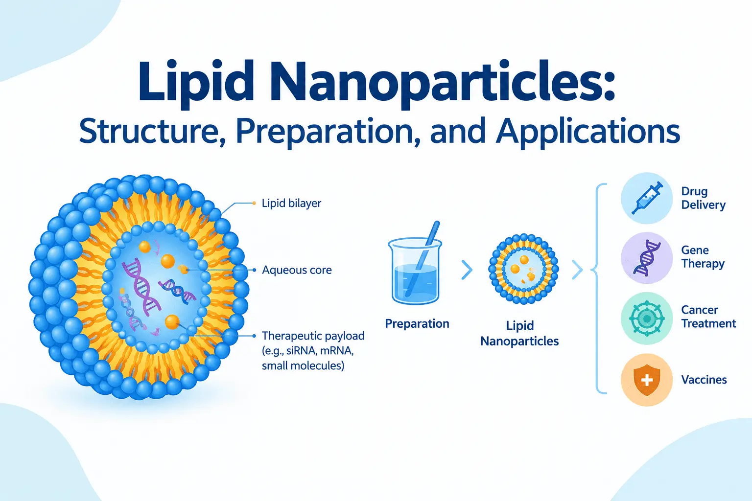
Lipid nanoparticles (LNPs) are nanoscale delivery systems widely used in biotechnology, molecular biology, and pharmaceutical research. They are particularly important for delivering nucleic acids, including messenger RNA (mRNA) and small interfering RNA (siRNA), into cells. By protecting these molecules from degradation and facilitating their intracellular delivery, lipid nanoparticles have become essential tools in mRNA vaccine development, gene therapy, and functional genomics.
Understanding lipid nanoparticle structure, preparation methods, characterization techniques, and biological mechanisms is important for researchers working in molecular biology and biomedical sciences.
1. What Are Lipid Nanoparticles?
Lipid nanoparticles are nanoscale particles composed of lipids that encapsulate or associate with therapeutic molecules. They are designed to improve the stability, delivery, and cellular uptake of biological substances that would otherwise be difficult to transport into cells.
Nucleic acids such as mRNA and siRNA are negatively charged and susceptible to degradation by nucleases. Their direct delivery into cells is also limited by the plasma membrane and intracellular barriers. Lipid nanoparticles help overcome these challenges by packaging nucleic acids within a protective lipid-based structure.
Lipid nanoparticles typically have diameters ranging from approximately 50 to 150 nanometers, although their size depends on the formulation and preparation method.
Unlike many conventional liposomes, which generally have an aqueous core surrounded by a lipid bilayer, lipid nanoparticles used for nucleic acid delivery often possess a more complex internal organization. Their structure depends on the lipid composition, the encapsulated cargo, and the formulation conditions.
The main functions of lipid nanoparticles include:
- Protecting nucleic acids against enzymatic degradation.
- Improving the cellular uptake of biological molecules.
- Facilitating the intracellular delivery of therapeutic cargo.
- Supporting the delivery of mRNA, siRNA, and other nucleic acids.
- Improving the stability and handling of certain biological formulations.
These properties make lipid nanoparticles valuable tools for studying gene expression, regulating gene activity, and developing nucleic acid-based therapeutics.
2. Structure and Components of Lipid Nanoparticles
The performance of lipid nanoparticles depends on their lipid composition. Most established LNP formulations for nucleic acid delivery contain four major lipid components: ionizable lipids, phospholipids, cholesterol, and polyethylene glycol (PEG)-conjugated lipids.
Each component contributes to particle formation, stability, cargo encapsulation, or intracellular delivery.
Ionizable lipids
Ionizable lipids are the principal components responsible for interacting with nucleic acids in many LNP formulations.
These lipids can change their electrical charge depending on the surrounding pH. During formulation, acidic conditions promote protonation of the ionizable lipids, allowing them to interact with negatively charged nucleic acids and facilitate their encapsulation.
At physiological pH, many ionizable lipids are predominantly neutral or less positively charged. This property can reduce unwanted interactions with biological membranes and help improve tolerability compared with permanently cationic lipids.
Following cellular uptake, the acidic environment of endosomes can promote lipid protonation and interactions with endosomal membranes, contributing to endosomal escape.
Phospholipids
Phospholipids are amphipathic molecules containing hydrophilic and hydrophobic regions. They contribute to the structural organization and stability of lipid nanoparticles.
Depending on their chemical structure, phospholipids can influence particle morphology, membrane interactions, and the physical properties of the formulation.
Cholesterol
Cholesterol helps regulate lipid packing and contributes to the structural organization of lipid nanoparticles. Its concentration can influence particle stability, lipid organization, and interactions with cellular membranes.
The optimal cholesterol content depends on the formulation and intended application.
PEGylated lipids
PEGylated lipids consist of a lipid anchor linked to polyethylene glycol (PEG). They are commonly incorporated into LNP formulations to help control particle size and reduce aggregation during preparation and storage.
PEG can also reduce nonspecific interactions with biological components. However, excessive PEG-lipid content may interfere with cellular uptake or intracellular delivery, making formulation optimization important.
Summary of lipid nanoparticle components
| Component | Main function |
|---|---|
| Ionizable lipids | Nucleic acid encapsulation and contribution to endosomal escape |
| Phospholipids | Structural organization and stability |
| Cholesterol | Lipid packing and membrane interactions |
| PEGylated lipids | Control of particle size, aggregation, and surface interactions |
The relative proportions of these components determine important formulation properties, including particle size, encapsulation efficiency, stability, cellular uptake, and biological activity.
3. How Are Lipid Nanoparticles Prepared?
Lipid nanoparticle preparation involves combining lipid components with the biological cargo under controlled conditions to produce particles with suitable physicochemical properties.
The preparation method can influence particle size, size distribution, encapsulation efficiency, reproducibility, and delivery performance.
Ethanol injection and rapid mixing
One common approach involves dissolving the lipid components in ethanol and mixing them with an aqueous solution containing the nucleic acid.
When the two solutions mix, the change in solvent environment reduces lipid solubility and promotes self-assembly into nanoparticles. Under appropriate conditions, the nucleic acid becomes encapsulated within the resulting particles.
Rapid and controlled mixing is important because it influences particle formation and size distribution.
Microfluidic mixing
Microfluidic mixing is widely used in lipid nanoparticle research because it enables controlled mixing of small fluid volumes.
In this method, lipid-containing ethanol and an aqueous nucleic acid solution flow through microchannels. The rapid exchange of solvents promotes lipid self-assembly and nucleic acid encapsulation.
Important parameters include:
- Total flow rate.
- Flow-rate ratio between the aqueous and ethanol phases.
- Lipid composition and concentration.
- Nucleic acid concentration.
- Buffer pH and ionic strength.
- Temperature and subsequent purification conditions.
Optimizing these parameters helps researchers produce reproducible formulations and adjust particle characteristics for specific applications.
Purification and formulation adjustment
After particle formation, residual ethanol and unencapsulated nucleic acids may need to be removed. Common approaches include dialysis, ultrafiltration, and size-exclusion chromatography, depending on the formulation and experimental requirements.
The formulation may then undergo buffer exchange and concentration adjustment before storage or further analysis.
Characterization of lipid nanoparticles
Characterization is essential for confirming that the prepared nanoparticles have suitable properties.
Dynamic light scattering (DLS): Estimates the hydrodynamic diameter of particles and provides information about their size distribution. The polydispersity index (PDI) helps assess the breadth of this distribution.
Zeta potential: Estimates the electrical potential near the particle’s slipping plane. It provides information about surface electrokinetic properties and can help assess formulation behavior, although it does not independently establish stability or biological performance.
Encapsulation efficiency: Measures the proportion of the initial nucleic acid associated with or encapsulated within the nanoparticles, according to the analytical method used. Fluorescence-based assays or other validated analytical approaches can be used.
Morphological analysis: Transmission electron microscopy (TEM) or cryogenic electron microscopy (cryo-EM) can provide information about particle morphology and internal organization.
Functional assays: Cell-based experiments can assess whether the formulation successfully delivers its cargo and produces the intended biological effect.
No single characterization method provides a complete assessment of LNP quality. Combining physicochemical measurements with functional testing offers a more informative evaluation.
4. How Do Lipid Nanoparticles Deliver Nucleic Acids Into Cells?
The delivery of nucleic acids by lipid nanoparticles involves several biological steps. Successful delivery requires not only cellular uptake but also the release of the cargo into the appropriate intracellular compartment.
Step 1: Cellular uptake
After exposure to cells, lipid nanoparticles can interact with the plasma membrane and enter cells through endocytic pathways.
During endocytosis, the cell membrane surrounds the particles and forms intracellular vesicles called endosomes.
Cell type, particle composition, surface properties, and experimental conditions can all influence cellular uptake.
Step 2: Endosomal trafficking
Following internalization, lipid nanoparticles are transported through the endosomal system. Endosomes undergo changes in composition and acidity as they mature.
A major challenge in nucleic acid delivery is that particles can remain trapped within endosomes and eventually be transported to lysosomes, where their cargo may be degraded.
Step 3: Endosomal escape
For many LNP formulations, only a fraction of the internalized nucleic acid reaches the cytoplasm.
As the endosomal environment becomes acidic, ionizable lipids can become protonated. Their interactions with endosomal membrane components may promote membrane destabilization and facilitate cargo release into the cytoplasm.
Endosomal escape is often a major limiting factor in LNP delivery efficiency. A formulation that achieves high cellular uptake does not necessarily produce efficient intracellular delivery.
Step 4: Release and biological activity
Once the nucleic acid reaches the appropriate intracellular compartment, it can produce its intended effect.
The outcome depends on the type of cargo:
- mRNA: Delivered mRNA can be translated by cytoplasmic ribosomes to produce a target protein.
- siRNA: Delivered siRNA can be incorporated into the RNA-induced silencing complex (RISC), which helps recognize and degrade complementary messenger RNA.
- Other nucleic acids: Depending on their type, structure, and delivery system, they may regulate gene expression or support other molecular biology applications.
The final biological response depends on cargo integrity, intracellular release, target-cell characteristics, and the properties of the nucleic acid itself.
5. Applications of Lipid Nanoparticles in Biotechnology and Medicine
Lipid nanoparticles have become important platforms in biomedical research because they can deliver nucleic acids without requiring them to cross cellular membranes unaided.
mRNA vaccine development
One of the best-known applications of lipid nanoparticles is the delivery of mRNA in vaccines.
The nanoparticles protect the mRNA and facilitate its delivery into cells. Once released into the cytoplasm, the mRNA can be translated into a target antigen, which can stimulate an immune response.
This technology has been used in COVID-19 vaccines and continues to be investigated for vaccines targeting other infectious diseases and certain cancers.
Gene therapy and genetic engineering
Lipid nanoparticles are being investigated as nonviral delivery systems for nucleic acids used in gene therapy and genetic engineering.
Potential cargo includes mRNA encoding a therapeutic protein, small interfering RNA, and components of gene-editing systems.
For example, LNPs can deliver messenger RNA encoding a genome-editing enzyme together with a guide RNA. The editing machinery can then act on its target sequence if it reaches the appropriate cellular compartment.
The efficiency and safety of these approaches depend on delivery specificity, cargo properties, immune responses, and the possibility of unintended biological effects.
RNA interference and functional genomics
In molecular biology, lipid nanoparticles can be used to deliver siRNA to investigate gene function.
After successful delivery, siRNA can reduce the expression of a complementary target gene. Researchers can then study how this reduction affects cellular processes, signaling pathways, or disease-associated phenotypes.
This approach is useful in functional genomics, target validation, and experimental studies of gene regulation.
Cancer research and drug delivery
Lipid nanoparticles are being studied for delivering therapeutic molecules to tumors and other disease-associated tissues.
Potential applications include the delivery of siRNA targeting cancer-related genes, mRNA encoding therapeutic proteins, and components of gene-editing systems.
Researchers are also investigating formulations designed to improve tissue distribution and cellular targeting. However, achieving selective delivery to tumor cells remains challenging because nanoparticles can accumulate in different tissues and may be affected by biological barriers within the tumor microenvironment.
Advantages and limitations of lipid nanoparticles
Lipid nanoparticles offer several advantages:
- They can protect nucleic acids against degradation.
- Their composition can be adjusted for different delivery applications.
- They support the delivery of several types of biological cargo.
- Their preparation can be scaled using controlled mixing technologies.
- They provide a nonviral alternative to certain nucleic acid delivery systems.
Nevertheless, important limitations remain. These include incomplete endosomal escape, variable delivery efficiency between cell types, potential inflammatory or immune responses, formulation instability, and challenges in achieving selective tissue targeting.
Future research is focused on improving lipid composition, optimizing intracellular delivery, increasing tissue specificity, and developing more stable formulations.
Conclusion
Lipid nanoparticles are versatile delivery systems that have become central to modern biotechnology and molecular medicine. Their structure, based on combinations of ionizable lipids, phospholipids, cholesterol, and PEGylated lipids, enables the encapsulation and intracellular delivery of nucleic acids.
Techniques such as rapid mixing and microfluidic formulation allow researchers to prepare nanoparticles with controlled physicochemical properties. Characterization methods, including dynamic light scattering, zeta potential analysis, encapsulation-efficiency assays, and microscopy, help evaluate formulation quality.
From mRNA vaccines to gene silencing and cancer research, lipid nanoparticles continue to expand the possibilities of nucleic acid delivery. Understanding their preparation, characterization, and intracellular mechanisms is essential for researchers developing new biotechnology tools and therapeutic strategies.


