

1. What Is a Peripheral Blood Smear?
Definition
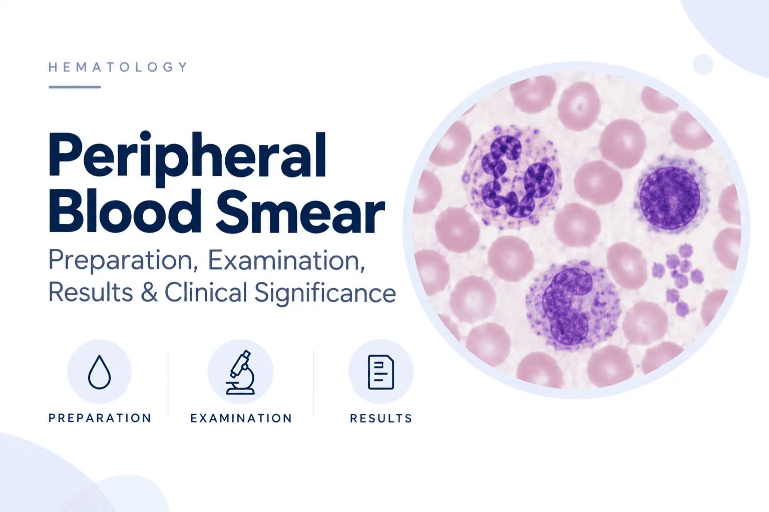
A peripheral blood smear, also called a peripheral blood film or simply a blood smear, is a microscopic preparation of peripheral blood.
A small drop of anticoagulated blood is spread into a thin film on a microscope slide. After drying and staining, the cells can be examined individually for their size, shape, color, nuclear characteristics, inclusions, and other morphological features.
The test can be performed as part of the investigation of an abnormal CBC results or when a clinician suspects a hematological disorder. It can also provide information that is difficult to obtain from cell counts alone.
Purpose of a Peripheral Blood Smear
The main purpose is to evaluate blood-cell morphology.
A smear can help answer questions such as:
- Are red blood cells unusually small or large?
- Are abnormal red-cell shapes present?
- Are white blood cells mature and morphologically normal?
- Are immature cells or blasts circulating in the blood?
- Do platelets appear adequate and normally distributed?
- Are blood parasites visible?
- Could an automated platelet count be falsely low because of platelet clumping?
Consequently, a smear is not simply another method of counting blood cells. Its major value is the additional morphological information it provides.
Peripheral Blood Smear vs. Complete Blood Count
A CBC, or complete blood count provides quantitative information such as hemoglobin concentration, hematocrit, RBC count, white-cell count, platelet count, MCV, and other calculated or measured parameters.
The smear provides a microscopic view of the cells themselves.
These tests therefore complement one another. For example, a CBC may reveal an increased MCV, while the smear can show whether the circulating red cells are predominantly macrocytic and whether other morphological abnormalities accompany the macrocytosis.
What Can Be Evaluated?
A properly prepared smear allows examination of three major cellular components:
- Red blood cells
- White blood cells
- Platelets
Depending on the clinical situation, the examiner may also look for microorganisms, abnormal inclusions, immature hematopoietic cells, and other unusual structures.
2. Components Examined in a Peripheral Blood Smear
Red Blood Cells
RBC examination focuses on several morphological characteristics:
- Size
- Shape
- Hemoglobinization
- Variation in size
- Variation in shape
- Intracellular inclusions
- Distribution and arrangement
Normal erythrocytes are approximately 7.5 μm in diameter, biconcave, and normally display a central area of pallor because of their shape and hemoglobin distribution.
Changes in these characteristics can help narrow the differential diagnosis of anemia and other hematological conditions.
White Blood Cells
The smear allows identification of the major circulating leukocyte populations:
- Neutrophils
- Lymphocytes
- Monocytes
- Eosinophils
- Basophils
Their relative proportions can be estimated manually, while their morphology can be examined for reactive changes, toxic changes, abnormal nuclear segmentation, immature forms, or malignant-appearing cells.
The identification of circulating blasts, for example, can be an important finding requiring urgent further investigation.
Platelets
Platelets are assessed for their approximate number, distribution, size, and appearance.
Microscopic examination can be particularly useful when an automated analyzer reports thrombocytopenia. Platelet clumping on the smear can indicate pseudothrombocytopenia, in which the measured platelet count is falsely low because platelets aggregate during specimen processing.
3. How Is a Peripheral Blood Smear Prepared?
Blood Sample Collection
Peripheral blood is commonly collected into an EDTA tube, which prevents coagulation while preserving cellular morphology.
The specimen should be properly mixed and transported promptly. Delays can produce degenerative changes and may alter the appearance or distribution of blood cells.
Preparing the Blood Film
The traditional method is the wedge or push technique.
A small drop of blood is placed near one end of a clean glass slide. A second slide, used as a spreader, is positioned in front of the drop and then pushed forward at an appropriate angle.
This produces a thin film that gradually becomes thinner toward the feathered edge.
A well-made smear should have a relatively smooth appearance and an appropriate area where RBCs are sufficiently separated for morphological assessment.
Air-Drying the Smear
After spreading, the film is allowed to air-dry.
Rapid and appropriate drying helps preserve cellular morphology. Excessive delay or inappropriate environmental conditions can contribute to artifacts that complicate interpretation.
Fixation and Staining
Once dried, the smear can be fixed and stained.
Commonly used stains include Wright, Wright-Giemsa, May-Grünwald-Giemsa, and other Romanowsky-type stains. These staining systems contain combinations of acidic and basic dyes that produce contrasting colors in different cellular structures.
For example, nuclei generally appear blue-purple, while hemoglobin-containing RBCs appear pink to reddish.
Quality Requirements for a Good Blood Smear
Smear quality directly affects interpretation.
A usable film should have:
- An even distribution of cells
- A gradual transition toward the feathered edge
- Adequate staining
- Minimal staining precipitate
- No excessive streaking or scratches
- A suitable monolayer for microscopic examination
Poor technique can create artifacts that mimic genuine abnormalities. For example, staining conditions can produce abnormal RBC shapes that are not truly present in the patient’s circulation.
4. How Is a Peripheral Blood Smear Examined?
Low-Power Examination
The examination begins at lower magnification to assess the overall quality and distribution of the smear.
The examiner identifies the region where cells are appropriately distributed and determines whether the film is suitable for detailed morphological evaluation.
The feathered edge may also be inspected because abnormal cells can sometimes become concentrated there.
Identifying the Monolayer
The monolayer is the region where RBCs are distributed sufficiently apart to allow their morphology to be assessed accurately.
If cells are examined in an excessively thick portion of the smear, they overlap and their shape can become difficult to evaluate. Conversely, cells at the extreme edge may appear distorted.
For this reason, selecting the appropriate examination area is an important part of smear interpretation.
High-Power Examination
Higher magnification is then used to examine individual cells.
Depending on the laboratory protocol, an oil-immersion objective may be used for detailed assessment of cellular morphology and differential leukocyte evaluation.
Manual Differential Leukocyte Count
A blood smear can also be used to perform a manual differential count, in which a defined number of leukocytes are classified according to their morphological appearance.
This is particularly useful when automated analyzers produce abnormal flags or when unusual cells require confirmation.
5. Red Blood Cell Morphology
RBC morphology is one of the most informative aspects of blood-film examination.
RBC Size
Red cells can be classified according to size:
- Microcytes — smaller than normal
- Normocytes — approximately normal size
- Macrocytes — larger than normal
Microcytosis can occur in iron deficiency and thalassemia, whereas macrocytosis may be associated with megaloblastic processes, alcohol exposure, liver disease, medications, and other conditions.
The MCV obtained from the CBC provides a quantitative measure of average RBC volume, while the smear shows the actual cellular appearance.
RBC Shape Abnormalities
Abnormally shaped RBCs are collectively described as poikilocytes.
Examples include:
- Spherocytes — dense, round RBCs lacking the usual central pallor
- Target cells — cells with a characteristic target-like appearance
- Schistocytes — fragmented RBCs
- Elliptocytes — elongated or elliptical cells
- Teardrop cells — pear-shaped RBCs
- Sickle cells — elongated, crescent-shaped RBCs
- Acanthocytes — irregularly spiculated RBCs
- Stomatocytes — RBCs with a slit-like central pallor
The significance of these forms depends on their quantity and the accompanying laboratory and clinical findings. For example, schistocytes can indicate RBC fragmentation and may occur in microangiopathic hemolysis.
RBC Color and Hemoglobinization
The amount and distribution of hemoglobin influence RBC coloration.
Hypochromia refers to increased central pallor and is commonly associated with reduced hemoglobinization.
Polychromasia describes RBCs that appear bluish-gray because relatively young red cells are circulating.
RBC Inclusions
Some RBCs contain visible intracellular structures that can provide additional diagnostic clues.
Examples include:
- Howell-Jolly bodies
- Basophilic stippling
- Pappenheimer bodies
- Heinz bodies
Their identification may suggest particular disorders of erythropoiesis, hemoglobin, red-cell metabolism, or splenic function.
6. White Blood Cell Morphology
White-cell examination goes beyond simply determining whether the WBC count is high or low.
Neutrophil Abnormalities
Neutrophils may show changes in nuclear segmentation and cytoplasmic appearance.
Hypersegmented neutrophils are characterized by excessive nuclear segmentation and are classically associated with megaloblastic hematopoiesis.
Other findings include toxic granulation, Döhle bodies, and cytoplasmic vacuolization, which can occur in reactive states such as severe infection or inflammation.
Lymphocyte Abnormalities
Reactive lymphocytes may become larger and develop more abundant cytoplasm. Such cells can occur during immune responses, particularly certain viral infections.
The identification of markedly atypical or abnormal lymphoid populations can also raise suspicion for a lymphoproliferative disorder and may require additional testing.
Monocyte, Eosinophil, and Basophil Abnormalities
Changes in monocyte morphology may occur in inflammatory and hematological disorders.
An increased number of eosinophils can accompany allergic disorders, certain drug reactions, and parasitic infections. Basophil abnormalities can also provide clues to particular myeloproliferative conditions.
Morphology and cell count should always be interpreted together rather than treating one abnormal cell type as diagnostic by itself.
Immature White Blood Cells
The appearance of immature granulocytes or blasts in peripheral blood can be clinically significant.
Blasts are immature hematopoietic cells that are normally found primarily in the bone marrow. A substantial circulating blast population can indicate an acute leukemia and requires urgent hematological evaluation.
7. Platelet Morphology
Platelet evaluation provides information that is not always available from the numerical platelet count alone.
Normal Platelet Appearance
Normal platelets are small, granular cytoplasmic fragments rather than complete cells with nuclei.
On a well-prepared smear, they should appear relatively evenly distributed without extensive aggregation.
Large and Giant Platelets
Some disorders are associated with unusually large platelets.
The presence of frequent giant platelets can occur in certain inherited thrombocytopenias and other hematological disorders. Their presence may also influence automated platelet counting because very large platelets can be misclassified by some analyzers.
Platelet Clumping
Platelet aggregates can cause an automated analyzer to report an unexpectedly low platelet count.
If the smear demonstrates extensive platelet clumping, the result may represent pseudothrombocytopenia rather than true thrombocytopenia. This distinction is clinically important because unnecessary investigations or treatment can otherwise follow a misleading laboratory result.
8. Common Findings on a Peripheral Blood Smear
A peripheral smear can provide clues across a broad range of conditions.
Findings Suggestive of Anemia
Depending on the cause, the smear may demonstrate microcytosis, macrocytosis, hypochromia, target cells, spherocytes, sickle cells, schistocytes, or other abnormalities.
These findings help classify the anemia and guide subsequent laboratory testing.
Findings Associated With Infection
Reactive leukocyte changes may accompany infection. Certain infections can also produce characteristic blood-cell patterns.
Most importantly, a smear can directly demonstrate some infectious organisms in blood.
Findings Suggestive of Hemolysis
RBC fragmentation, spherocytes, bite cells, or other forms of cellular injury may provide evidence supporting hemolysis and help distinguish possible mechanisms.
Findings Associated With Leukemia
The presence of blasts or an abnormal population of leukocytes can raise suspicion for leukemia. However, microscopic morphology is generally the starting point for further characterization rather than the final diagnostic test.
Flow cytometry, cytogenetic studies, molecular testing, and bone-marrow examination may be required depending on the findings.
Findings Associated With Parasitic Infections
Some blood parasites can be directly visualized on a peripheral smear.
Plasmodium, the parasite responsible for malaria, is an important example. Examination of appropriately prepared and stained blood films can identify infected RBCs and provide morphological information useful for parasite identification.
11. Advantages and Limitations of Peripheral Blood Smear Examination
Advantages
Peripheral blood smear examination offers several important benefits:
- Direct visualization of individual blood cells
- Assessment of cellular morphology
- Detection of abnormal or immature cells
- Recognition of some blood parasites
- Identification of platelet clumping
- Confirmation of certain abnormalities flagged by automated analyzers
- Relatively simple laboratory methodology
It can therefore remain highly informative even in laboratories equipped with modern automated hematology systems.
Limitations
The technique also has limitations.
Interpretation requires appropriate training and experience. Results can be affected by specimen quality, smear preparation, staining, storage, and technical artifacts.
In addition, morphology alone may not distinguish between conditions with similar appearances. Some diagnoses require immunophenotyping, biochemical testing, cytogenetics, molecular testing, or bone-marrow examination.
Thus, the value of a blood smear depends not only on microscopy but also on proper clinical and laboratory correlation.
12. Peripheral Blood Smear vs. Automated Hematology Analyzer
Modern hematology analyzers can rapidly measure blood-cell numbers and provide numerous indices and automated differential counts.
However, automated analysis does not eliminate the need for microscopic examination.
A smear can provide information that an analyzer may not fully characterize, particularly when abnormal cell morphology or unusual populations are present. Automated instruments may flag suspected abnormalities, prompting a laboratory professional to review the blood film manually.
The two approaches are therefore complementary:
| Automated CBC | Peripheral blood smear |
|---|---|
| Primarily quantitative | Primarily morphological |
| Rapid, high-throughput analysis | Detailed microscopic examination |
| Provides cell counts and indices | Shows individual cell appearance |
| Automated differential | Manual morphological assessment |
| Can generate instrument flags | Can investigate flagged abnormalities |
The most informative hematological evaluation often combines both methods rather than treating them as competing tests.
Medical Disclaimer: This content is for informational and educational purposes only and does not constitute medical advice. It is not intended to replace professional medical consultation, diagnosis, or treatment. Always seek the advice of a qualified healthcare provider with any questions regarding a medical condition.
References and Further Reading
- Mohammed EA, Mohamed MM, Far BH, Naugler C. Peripheral blood smear image analysis: A comprehensive review. J Pathol Inform. 2014 Mar 28;5(1):9. doi: 10.4103/2153-3539.129442.
- Kaferle J, Strzoda CE. Evaluation of macrocytosis. Am Fam Physician. 2009 Feb 1;79(3):203-8. PMID: 19202968.
- Chabot-Richards DS, George TI. Leukocytosis. Int J Lab Hematol. 2014 Jun;36(3):279-88. doi: 10.1111/ijlh.12212.
- Chase ML, Drews R, Zumberg MS, Ellis LR, Reid EG, Gerds AT, Lee AI, Hobbs GS, Berry J, Freed JA. Consensus recommendations on peripheral blood smear review: defining curricular standards and fellow competency. Blood Adv. 2023 Jul 11;7(13):3244-3252. doi: 10.1182/bloodadvances.2023009843.


