

Key Takeaways
- Angiogenesis is the process of forming new blood vessels from pre-existing ones.
- It plays an essential role in embryonic development by supplying growing tissues with oxygen and nutrients.
- Angiogenesis supports wound healing and tissue repair by restoring blood supply to damaged areas.
- It contributes to normal physiological processes, including the menstrual cycle, pregnancy, and muscle adaptation to exercise.
- The process involves several steps, including endothelial cell activation, migration, proliferation, and the formation of new vessel structures.
- VEGF (vascular endothelial growth factor) is a major regulator that stimulates endothelial cell growth and migration.
- Other signaling molecules, such as FGF, angiopoietins, and the Notch pathway, help control vessel formation and maturation.
- Hypoxia, or low oxygen availability, can stimulate angiogenesis through the activation of HIF-1 signaling.
- Abnormal angiogenesis is associated with several diseases, including cancer, diabetic retinopathy, age-related macular degeneration, and chronic inflammatory disorders.
- Anti-angiogenic therapies can inhibit excessive blood vessel formation, while pro-angiogenic approaches aim to improve blood supply in ischemic tissues.
1. What Is Angiogenesis?
Definition and Biological Importance of Angiogenesis
Angiogenesis is the formation of new blood vessels from an existing vascular network. It primarily involves endothelial cells, which line the inner surface of blood vessels and respond to signals that stimulate their migration, proliferation, and organization into new vascular structures.
This process allows the vascular system to expand and adapt to the metabolic requirements of different tissues. For example, growing tissues require additional blood vessels to maintain adequate oxygen and nutrient delivery.
Angiogenesis is regulated by a balance between pro-angiogenic and anti-angiogenic molecules. This balance ensures that new vessels form when necessary and that vascular growth is controlled.
Angiogenesis vs. Vasculogenesis and Arteriogenesis
Although angiogenesis, vasculogenesis, and arteriogenesis all contribute to blood vessel development, they describe different biological processes.
- Angiogenesis: Formation of new blood vessels from pre-existing vessels through endothelial cell proliferation and migration.
- Vasculogenesis: Formation of blood vessels from endothelial progenitor cells or angioblasts, particularly during embryonic development.
- Arteriogenesis: Enlargement and remodeling of pre-existing small arteries or collateral vessels, often in response to increased blood flow and shear stress.
These mechanisms work together to establish and maintain an efficient vascular network.
Physiological vs. Pathological Angiogenesis
Angiogenesis can be classified into two major forms:
Physiological angiogenesis occurs under normal conditions, including embryonic development, wound healing, pregnancy, and tissue adaptation to exercise.
Pathological angiogenesis occurs when vascular growth becomes excessive, insufficient, or improperly regulated. It contributes to diseases such as cancer, diabetic retinopathy, and chronic inflammatory disorders.
The difference between these two forms depends largely on the balance of angiogenic signals and the local tissue environment.
2. Mechanisms and Steps of Angiogenesis
Angiogenesis involves several coordinated cellular and molecular events. Although different angiogenic mechanisms exist, sprouting angiogenesis is one of the best-characterized forms.
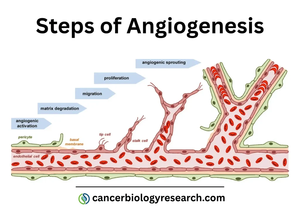
Adapted from: Laschke MW, Gu Y and Menger MD (2022) Replacement in angiogenesis research: Studying mechanisms of blood vessel development by animal-free in vitro, in vivo and in silico approaches. Front. Physiol. 13:981161. https://doi.org/10.3389/fphys.2022.981161
Activation of Endothelial Cells
Angiogenesis usually begins when tissues experience conditions that stimulate blood vessel formation, such as hypoxia (low oxygen availability).
Hypoxic cells release pro-angiogenic factors, particularly vascular endothelial growth factor (VEGF). VEGF binds to receptors on endothelial cells, activating intracellular signaling pathways that promote their survival, migration, and proliferation.
This activation causes endothelial cells to become more responsive to angiogenic signals.
Degradation of the Basement Membrane and Extracellular Matrix
To form new vascular sprouts, endothelial cells must pass through the basement membrane and surrounding extracellular matrix (ECM).
Matrix metalloproteinases (MMPs), particularly MMP-2 and MMP-9, help degrade extracellular matrix components and facilitate endothelial cell migration.
This remodeling creates space for endothelial cells to move toward angiogenic signals.
Endothelial Cell Migration and Proliferation
Following activation, endothelial cells migrate toward the angiogenic stimulus.
Two important endothelial cell populations participate in sprouting angiogenesis:
- Tip cells: Specialized endothelial cells located at the leading edge of a growing vascular sprout. They guide migration by extending filopodia and sensing angiogenic signals.
- Stalk cells: Endothelial cells located behind tip cells. They proliferate and contribute to the elongation of the developing vessel.
The coordination between these cells allows vascular sprouts to extend into surrounding tissues.
Formation of New Vascular Tubes and Lumen
As endothelial cells migrate and proliferate, they organize into tubular structures.
These cells establish cell-to-cell connections and develop a central lumen through which blood can eventually flow.
When two growing vascular sprouts meet, they can connect through a process called anastomosis, establishing continuity between the newly formed vessels and the existing circulation.
Vessel Maturation and Stabilization
Newly formed blood vessels are initially immature and require additional stabilization.
Pericytes and vascular smooth muscle cells are recruited to support endothelial cells and regulate vascular integrity.
The basement membrane is also reorganized, and endothelial cell junctions become more stable.
These changes produce functional blood vessels capable of maintaining blood flow and regulating vascular permeability.
3. Key Factors and Signaling Pathways Regulating Angiogenesis
Angiogenesis is controlled by a complex network of growth factors, receptors, and intracellular signaling pathways. These molecular regulators determine when and where new blood vessels develop.
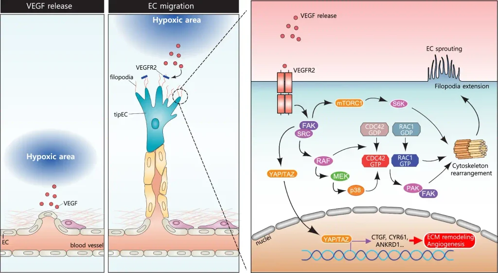
Source: Shah, F.H., Nam, Y.S., Bang, J.Y. et al. Targeting vascular endothelial growth receptor-2 (VEGFR-2): structural biology, functional insights, and therapeutic resistance. Arch. Pharm. Res. 48, 404–425 (2025). https://doi.org/10.1007/s12272-025-01545-1
Vascular Endothelial Growth Factor (VEGF)
Vascular endothelial growth factor (VEGF) is one of the most important regulators of angiogenesis.
The VEGF family includes several members, including VEGF-A, VEGF-B, VEGF-C, and VEGF-D. VEGF-A is particularly important in blood vessel formation.
VEGF-A binds primarily to VEGFR-2 receptors on endothelial cells, activating signaling pathways that stimulate:
- Endothelial cell proliferation and survival.
- Migration toward angiogenic stimuli.
- Increased vascular permeability.
- Formation of new vascular sprouts.
VEGF production is strongly influenced by hypoxia through hypoxia-inducible factor 1 (HIF-1).
Fibroblast Growth Factors (FGFs)
Fibroblast growth factors are another important group of proteins involved in angiogenesis.
FGF-2, also known as basic fibroblast growth factor (bFGF), promotes endothelial cell proliferation, migration, and survival.
FGFs also stimulate the production of other angiogenic mediators and contribute to extracellular matrix remodeling.
These factors participate in tissue development, wound healing, and vascular repair.
Angiopoietins and Tie Receptors
Angiopoietins regulate blood vessel maturation and stability through their interaction with Tie receptors expressed on endothelial cells.
Two major members of this family are:
- Angiopoietin-1 (Ang-1): Promotes vascular stabilization, endothelial cell survival, and vessel integrity through Tie2 activation.
- Angiopoietin-2 (Ang-2): Can destabilize existing vessels and facilitate vascular remodeling, particularly in the presence of VEGF.
The balance between Ang-1 and Ang-2 is important for controlling vascular remodeling and angiogenesis.
Notch and Delta-Like Ligand 4 (DLL4) Signaling
The Notch signaling pathway plays an important role in coordinating endothelial cell behavior during sprouting angiogenesis.
DLL4 is expressed by endothelial tip cells and activates Notch receptors in neighboring endothelial cells.
This signaling helps regulate tip and stalk cell differentiation, preventing excessive formation of competing vascular sprouts.
Consequently, DLL4-Notch signaling contributes to the organization and patterning of the developing vascular network.
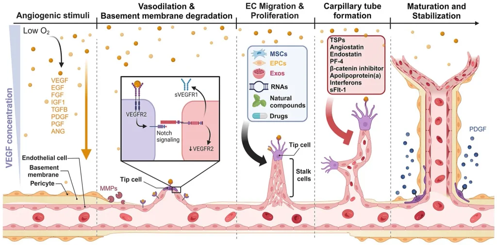
Source: Zhu W, Xu Z, Zhou D, Xu J, He Y, Li ZA. Bioengineering strategies targeting angiogenesis: Innovative solutions for osteonecrosis of the femoral head. Journal of Tissue Engineering. 2025;16. https://doi.org/10.1177/20417314241310541
Hypoxia-Inducible Factor 1 (HIF-1)
Hypoxia-inducible factor 1 (HIF-1) is a transcription factor that regulates cellular responses to low oxygen levels.
Under normal oxygen conditions, the HIF-1α subunit is hydroxylated and targeted for degradation through the von Hippel–Lindau (VHL) protein pathway.
Under hypoxic conditions, HIF-1α becomes stabilized, enters the nucleus, and forms a complex with HIF-1β.
This complex activates the expression of several genes involved in adaptation to hypoxia, including VEGF.
HIF-1 therefore provides an important molecular link between tissue oxygen availability and angiogenesis.
Angiogenesis Inhibitors and Endogenous Regulation
Angiogenesis is also controlled by endogenous inhibitors that prevent uncontrolled vascular growth.
Important anti-angiogenic molecules include:
- Thrombospondin-1: Inhibits endothelial cell proliferation and migration.
- Endostatin: A fragment derived from collagen XVIII that suppresses angiogenic activity.
- Angiostatin: A plasminogen-derived fragment with anti-angiogenic properties.
The interaction between these inhibitors and pro-angiogenic factors maintains vascular homeostasis.
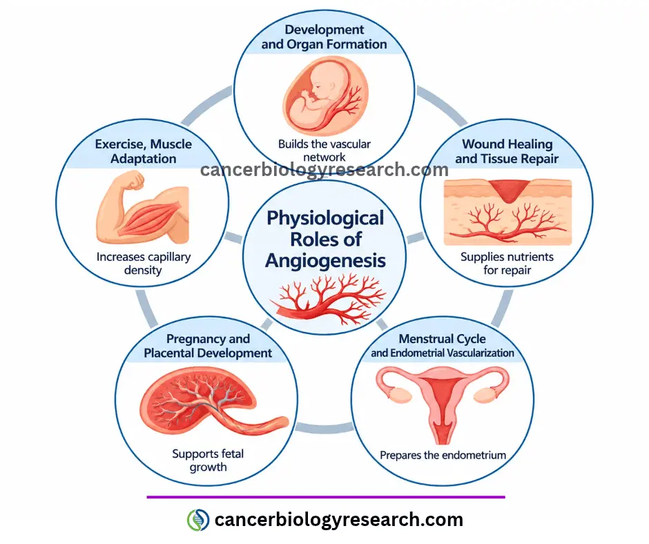
4. Physiological Roles of Angiogenesis in the Human Body
Angiogenesis is essential for several normal physiological processes throughout life. It allows tissues to develop, repair damage, and adapt to changes in metabolic requirements.
Embryonic Development and Organ Formation
During embryonic development, angiogenesis contributes to the formation of the vascular network required for organ growth and function.
As tissues develop, their metabolic requirements increase, making an efficient blood supply essential.
Angiogenesis allows existing vessels to expand and form new branches, supporting the development of organs and tissues.
It also contributes to the establishment of vascular networks that connect different regions of the developing organism.
Wound Healing and Tissue Repair
Angiogenesis is a fundamental component of wound healing.
Following tissue injury, inflammatory cells and damaged tissues release growth factors and cytokines that stimulate endothelial cell activation.
New blood vessels develop within the healing tissue, providing oxygen, nutrients, and immune cells required for tissue repair.
VEGF is particularly important during this process because it stimulates endothelial cell migration and proliferation.
As healing progresses, newly formed vessels undergo maturation and remodeling to support tissue regeneration.
Menstrual Cycle and Endometrial Vascularization
The endometrium, the inner lining of the uterus, undergoes continuous structural changes during the menstrual cycle.
Angiogenesis contributes to the development and remodeling of endometrial blood vessels, particularly during the proliferative and secretory phases.
These vascular changes help prepare the endometrium for potential embryo implantation.
If pregnancy does not occur, hormonal changes trigger menstruation and tissue shedding, followed by subsequent regeneration.
Pregnancy and Placental Development
Angiogenesis is essential for the development of the placenta, which provides oxygen and nutrients to the developing fetus.
During placental development, new blood vessels form within the fetal component of the placenta, supporting the establishment of an efficient maternal-fetal exchange system.
VEGF and placental growth factor (PlGF) are important regulators of placental vascular development.
Abnormal regulation of angiogenesis can contribute to pregnancy complications, including preeclampsia and placental insufficiency.
Exercise, Muscle Adaptation, and Tissue Oxygenation
Physical exercise increases the metabolic demands of skeletal muscles.
In response to repeated exercise, particularly endurance training, skeletal muscle can undergo vascular adaptations that increase capillary density.
This process improves oxygen and nutrient delivery while supporting the removal of metabolic waste products.
Exercise-induced angiogenesis is influenced by several factors, including VEGF, mechanical signals, and changes in tissue oxygen availability.
These adaptations contribute to improved muscle endurance and metabolic efficiency.
5. Angiogenesis in Diseases and Clinical Applications
Although angiogenesis is essential for normal physiology, abnormal regulation of this process contributes to several pathological conditions. Depending on the disease, excessive or insufficient blood vessel formation can interfere with normal tissue function.
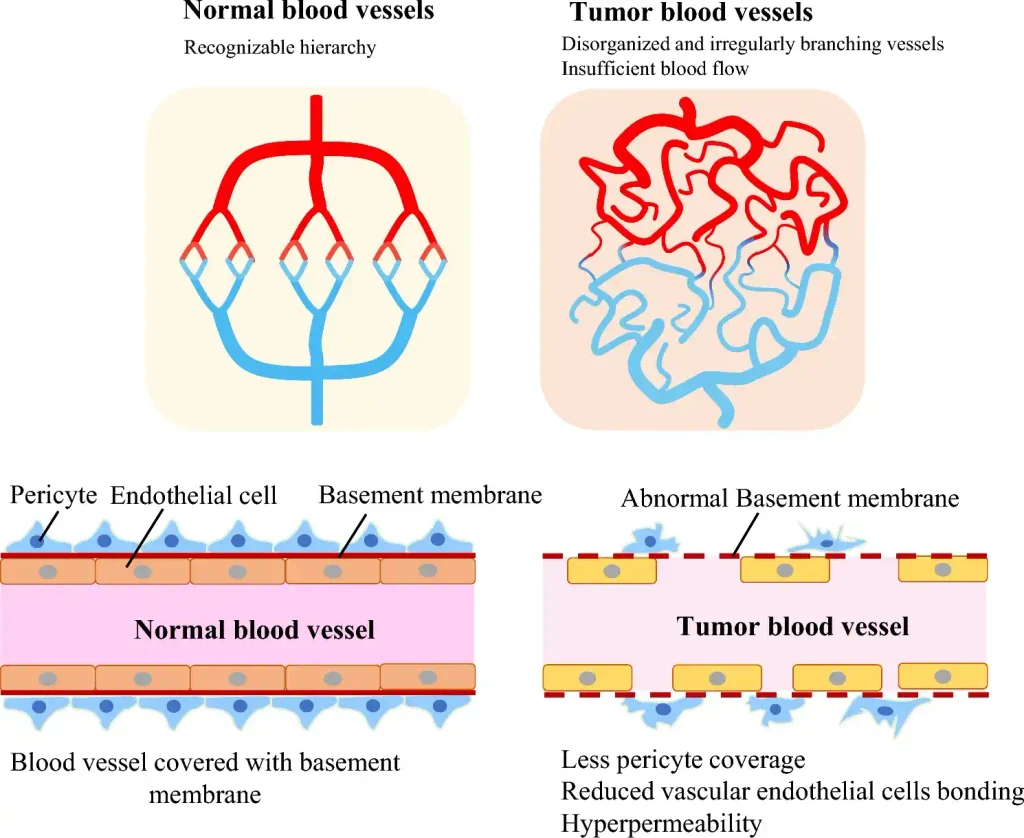
Source: Hida, K., Maishi, N., Matsuda, A. et al. Beyond starving cancer: anti-angiogenic therapy. J Med Ultrasonics 51, 605–610 (2024). https://doi.org/10.1007/s10396-023-01310-1
Angiogenesis in Cancer and Tumor Progression
Cancer cells require oxygen and nutrients to support continuous growth. As tumors enlarge, they can experience hypoxia, which stimulates the production of angiogenic factors such as VEGF.
This promotes the formation of new blood vessels that supply the tumor with nutrients and oxygen.
However, tumor-associated blood vessels are often structurally abnormal, with irregular branching, increased permeability, and inefficient blood flow.
These abnormal vessels can also contribute to tumor progression, immune suppression, and metastatic dissemination.
For a more detailed explanation, see our article on Angiogenesis in Cancer.
Diabetic Retinopathy and Age-Related Macular Degeneration
Abnormal angiogenesis contributes to several retinal diseases that can cause vision impairment.
In diabetic retinopathy, chronic hyperglycemia damages retinal blood vessels. Advanced disease can lead to retinal ischemia, which stimulates VEGF production and the formation of abnormal new vessels.
These fragile vessels can bleed and cause retinal damage.
In neovascular age-related macular degeneration (AMD), abnormal blood vessels develop beneath or within the retina, potentially causing fluid leakage, bleeding, and progressive vision loss.
Anti-VEGF treatments are widely used to control abnormal vascular growth in these conditions.
Chronic Inflammation and Rheumatoid Arthritis
Angiogenesis also participates in chronic inflammatory diseases.
In rheumatoid arthritis, inflammation of the synovial membrane stimulates the production of VEGF and other pro-angiogenic factors.
New blood vessels develop within the inflamed synovial tissue, helping supply inflammatory cells and supporting the expansion of the abnormal tissue.
This process contributes to persistent inflammation and joint damage.
Angiogenesis therefore represents one component of the complex interaction between vascular development and immune responses.
Ischemic Diseases and Cardiovascular Disorders
Insufficient blood supply can cause tissue ischemia, which occurs when oxygen delivery does not meet metabolic requirements.
In conditions such as coronary artery disease and peripheral artery disease, restricted blood flow can damage tissues and impair organ function.
Angiogenesis represents an adaptive response to ischemia by promoting the formation of new blood vessels.
Therapeutic strategies that stimulate vascular growth are being investigated to improve blood supply in ischemic tissues.
However, successful treatment requires the development of functional vessels capable of providing effective tissue perfusion.
Angiogenesis as a Therapeutic Target
Because angiogenesis contributes to several diseases, regulating this process has become an important therapeutic strategy.
Two major approaches are used:
Anti-angiogenic therapy
These treatments inhibit excessive blood vessel formation. They are particularly important in cancer and retinal diseases.
Examples include VEGF-targeting drugs such as bevacizumab, ranibizumab, and aflibercept.
Pro-angiogenic therapy
This approach aims to stimulate blood vessel formation in tissues affected by insufficient vascularization.
Potential applications include ischemic cardiovascular diseases, peripheral artery disease, and tissue repair.
Strategies involving growth factors, cell-based therapies, and tissue engineering are being investigated to improve vascular regeneration.
However, therapeutic angiogenesis remains challenging because newly formed vessels must be stable, properly organized, and functionally connected to the existing circulation.
Conclusion
Angiogenesis is a fundamental physiological process responsible for the formation of new blood vessels from pre-existing vascular structures. It plays essential roles in embryonic development, wound healing, pregnancy, tissue repair, and adaptation to physical exercise.
This process is regulated by a complex network of molecular factors, including VEGF, FGFs, angiopoietins, HIF-1, and Notch signaling. Their coordinated activity ensures that blood vessel formation occurs according to tissue requirements.
However, abnormal angiogenesis contributes to several diseases, including cancer, diabetic retinopathy, age-related macular degeneration, and chronic inflammatory disorders. Understanding its molecular mechanisms has therefore opened important opportunities for developing therapies that either inhibit excessive vascular growth or stimulate vascular regeneration.
Overall, angiogenesis represents an essential link between vascular biology, tissue physiology, and disease development.
References and Further Reading
- Laschke MW, Gu Y and Menger MD (2022) Replacement in angiogenesis research: Studying mechanisms of blood vessel development by animal-free in vitro, in vivo and in silico approaches. Front. Physiol. 13:981161.https://doi.org/10.3389/fphys.2022.981161
- Shah, F.H., Nam, Y.S., Bang, J.Y. et al. Targeting vascular endothelial growth receptor-2 (VEGFR-2): structural biology, functional insights, and therapeutic resistance. Arch. Pharm. Res. 48, 404–425 (2025). https://doi.org/10.1007/s12272-025-01545-1
- Zhu W, Xu Z, Zhou D, Xu J, He Y, Li ZA. Bioengineering strategies targeting angiogenesis: Innovative solutions for osteonecrosis of the femoral head. Journal of Tissue Engineering. 2025;16. https://doi.org/10.1177/20417314241310541
- Hida, K., Maishi, N., Matsuda, A. et al. Beyond starving cancer: anti-angiogenic therapy. J Med Ultrasonics 51, 605–610 (2024). https://doi.org/10.1007/s10396-023-01310-1
- Potente M, Gerhardt H, Carmeliet P. (2011). Basic and therapeutic aspects of angiogenesis. Cell, 146(6), 873–887. https://doi.org/10.1016/j.cell.2011.08.039
- Carmeliet P, Jain RK. (2011). Molecular mechanisms and clinical applications of angiogenesis. Nature, 473, 298–307. https://doi.org/10.1038/nature10144
- Favier J, Corvol P. (2001). Physiological angiogenesis. The Journal of Physiology, 536, 455–463. https://doi.org/10.1111/j.1469-7793.2001.tb00688.x


