

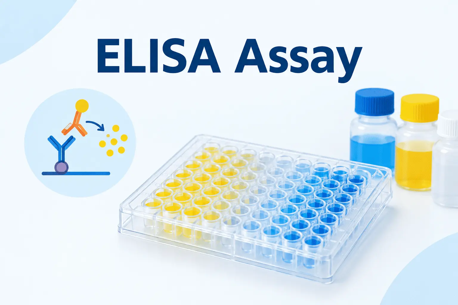
The enzyme-linked immunosorbent assay (ELISA) is a widely used laboratory technique for detecting and measuring specific antigens or antibodies in biological samples. The ELISA test combines the specificity of antigen–antibody interactions with an enzyme-mediated reaction that produces a measurable signal, usually a color change.
Because of its sensitivity, specificity, and ability to analyze multiple samples simultaneously, the ELISA assay is widely used in medical diagnostics, immunology, microbiology, pharmaceutical research, and biomarker studies.
In this guide, you will learn the ELISA principle, the four main types of ELISA, the step-by-step procedure, how to interpret ELISA test results, and the advantages and limitations of this technique.
What Is an ELISA Test?
ELISA stands for enzyme-linked immunosorbent assay. It is an immunological technique used to detect or quantify a target substance, known as an analyte, through specific binding interactions between antigens and antibodies.
The target analyte may be a protein, antibody, hormone, cytokine, microbial antigen, or another substance for which a suitable assay has been developed.
An ELISA test is commonly performed in microplates containing multiple wells. These wells provide a solid surface on which antigens or antibodies can be immobilized. After the sample and detection reagents are added, an enzyme–substrate reaction generates a signal that can be measured using a microplate reader.
Depending on the assay design, ELISA can provide:
- Qualitative results: Indicating whether the target is detected.
- Semiquantitative results: Estimating the target level relative to a reference or cutoff.
- Quantitative results: Measuring the target concentration using a calibration curve.
The ELISA assay is particularly useful when a laboratory needs to analyze many samples under standardized conditions.
ELISA Principle
The ELISA principle relies on the specific interaction between an antigen and an antibody, combined with an enzyme-linked detection system.
An antigen is a substance that can be specifically recognized by an antibody. Antibodies, or immunoglobulins, bind to particular molecular structures called epitopes. ELISA uses this recognition process to capture or detect a target analyte.
The general ELISA principle involves four main elements:
- Specific recognition: An antibody binds to its target antigen, or an antigen binds to a target antibody.
- Immobilization: One assay component is attached to a solid surface, usually the bottom of a microplate well.
- Enzyme-linked detection: An enzyme conjugated to an antibody or another assay component helps generate a detectable signal.
- Signal measurement: A suitable substrate reacts with the enzyme to produce a measurable signal, often a color change.
Common enzymes used in ELISA include horseradish peroxidase (HRP) and alkaline phosphatase (AP). HRP can react with substrates such as tetramethylbenzidine (TMB), producing a colored product. In many TMB-based assays, adding an acidic stop solution changes the reaction product’s color, allowing absorbance to be measured at the wavelength specified by the assay protocol.
Relationship Between Color Intensity and Analyte Concentration
In many conventional ELISA formats, the measured signal increases with the concentration of the target analyte. However, this relationship depends on the assay design.
In a sandwich ELISA, for example, more captured antigen generally leads to more enzyme-linked detection antibody and a stronger signal within the validated measurement range.
In a competitive ELISA, the relationship is often reversed: a higher concentration of the target analyte produces a lower signal.
Therefore, ELISA results must be interpreted according to the specific assay format, calibration curve, and validated analytical range.
Types of ELISA Assays
There are four commonly described ELISA formats: direct ELISA, indirect ELISA, sandwich ELISA, and competitive ELISA. They differ in how the target analyte is captured and detected.
1. Direct ELISA
Direct ELISA detects an antigen using an enzyme-labeled antibody that binds directly to the target.
Procedure:
- Immobilize the antigen on the microplate surface.
- Block unoccupied binding sites.
- Add an enzyme-conjugated primary antibody specific to the antigen.
- Wash away unbound antibody.
- Add the enzyme substrate.
- Measure the resulting signal.
Advantages:
- Relatively simple procedure.
- Fewer incubation and detection steps.
- Reduced risk of nonspecific interactions involving secondary antibodies.
Limitations:
- Each primary antibody generally needs its own enzyme conjugate.
- Signal amplification may be limited compared with some indirect formats.
- Protein adsorption to the plate can affect antigen conformation and antibody recognition.
Direct ELISA is useful for detecting immobilized antigens when an appropriate labeled antibody is available.
2. Indirect ELISA
Indirect ELISA commonly detects antibodies in a biological sample, such as serum. It uses an antigen attached to the microplate and a labeled secondary antibody to detect antibodies that bind to that antigen.
Procedure:
- Coat the microplate with the relevant antigen.
- Block unoccupied binding sites.
- Add the patient’s serum or another antibody-containing sample.
- Wash away unbound sample components.
- Add an enzyme-conjugated secondary antibody that recognizes the target antibody.
- Wash and add the enzyme substrate.
- Measure the signal.
For example, an indirect ELISA may be designed to detect antibodies against a particular infectious agent.
Advantages:
- A single labeled secondary antibody can be used with multiple compatible primary antibodies.
- Multiple secondary antibodies may bind to a target antibody, potentially increasing signal.
- Suitable for antibody detection in many research and diagnostic applications.
Limitations:
- Cross-reactivity of the secondary antibody may produce nonspecific signals.
- Additional incubation and washing steps are required.
- A positive antibody result does not necessarily indicate an active infection; interpretation depends on the disease, antibody class, timing, and clinical context.
3. Sandwich ELISA
Sandwich ELISA detects an antigen using two antibodies that recognize different epitopes of the same target. The antigen is captured between a capture antibody and a detection antibody, forming a molecular complex that resembles a sandwich.
Procedure:
- Coat the microplate with a capture antibody.
- Block unoccupied binding sites.
- Add the sample containing the target antigen.
- Wash away unbound sample components.
- Add a detection antibody that binds to a different epitope of the antigen.
- Add an enzyme-linked detection reagent if the detection antibody is not already enzyme-labeled.
- Wash and add the substrate.
- Measure the resulting signal.
Sandwich ELISA is widely used to quantify proteins, cytokines, hormones, and other analytes in biological samples.
Advantages:
- Often provides high specificity because two recognition events are involved.
- Can achieve high sensitivity with a suitable antibody pair.
- Suitable for complex samples, including serum and plasma, when appropriately validated.
Limitations:
- Requires compatible capture and detection antibodies.
- Antibody development and assay optimization can be demanding.
- Very high antigen concentrations can sometimes produce a hook effect, resulting in unexpectedly low signals in certain sandwich assay designs.
Sandwich ELISA is particularly useful when the target is present at low concentrations or within a complex biological matrix.
4. Competitive ELISA
Competitive ELISA measures an analyte through competition between sample components and a reference or labeled component for limited binding sites.
The exact design varies. For example, a sample analyte may compete with a labeled analyte for antibody binding, or sample antigen may compete with an immobilized antigen for a limited amount of antibody.
In a common competitive format, a higher concentration of the target analyte results in less labeled component being retained, producing a lower signal.
General procedure:
- Prepare the assay’s immobilized binding component or antibody system.
- Introduce the sample and the appropriate competing reagent.
- Allow the competitive binding reaction to occur.
- Wash away unbound components.
- Add the substrate when an enzyme-linked component is used.
- Measure the signal and interpret it using the assay’s calibration curve.
Advantages:
- Useful for small molecules that may not accommodate two antibodies simultaneously.
- Can be adapted to hormones, drugs, toxins, and other analytes.
- Suitable for analytes for which a sandwich format is impractical.
Limitations:
- The signal–concentration relationship can be less intuitive.
- Assay optimization and calibration are important for accurate quantification.
- The interpretation depends strongly on the specific competitive design.
Comparison of the Four ELISA Types
| Feature | Direct ELISA | Indirect ELISA | Sandwich ELISA | Competitive ELISA |
|---|---|---|---|---|
| Typical target | Antigen | Antibody | Antigen | Antigen or other analyte |
| Main detection approach | Labeled primary antibody | Labeled secondary antibody | Capture and detection antibodies | Competitive binding |
| Number of recognition antibodies | Usually one | Primary and secondary | Usually two target-specific antibodies | Depends on the format |
| Signal with increasing analyte | Often increases | Often increases with target antibody | Usually increases | Often decreases |
| Common application | Antigen detection | Antibody detection | Protein quantification | Small-molecule measurement |
These are general patterns; individual commercial assays may use different configurations.
ELISA Test Procedure: Step by Step
Although exact protocols vary, most conventional ELISA assays follow a similar workflow. The procedure must be adapted to the selected kit, target analyte, sample type, and detection system.
Step 1: Prepare the Microplate
The wells of a microplate provide the solid phase for the assay. Depending on the ELISA format, the plate is coated with an antigen or capture antibody, or a pre-coated plate is supplied.
The coating conditions influence how effectively the target is captured and recognized.
Step 2: Block Unoccupied Binding Sites
After coating, some areas of the plate surface remain available for nonspecific protein adsorption. A blocking solution is added to occupy these sites and reduce background signal.
Common blocking agents include bovine serum albumin (BSA), casein, and other proteins or formulations compatible with the assay.
Step 3: Add the Sample
The biological sample is added to the wells. If the target analyte is present and accessible, it binds to the immobilized capture component or other assay reagent.
Samples may include serum, plasma, cell culture supernatants, tissue extracts, or other validated matrices.
Sample dilution and pretreatment should follow the assay protocol.
Step 4: Wash the Wells
Washing removes unbound substances while retaining the specifically bound components.
Insufficient washing can increase background signal, whereas excessively harsh washing may disrupt specific binding. Consistent washing is essential for reproducible results.
Step 5: Add the Detection Reagent
A detection antibody or other enzyme-linked reagent is introduced to recognize the target or an antibody bound during an earlier step.
In sandwich ELISA, the detection antibody generally recognizes a different epitope from the capture antibody. In indirect ELISA, an enzyme-linked secondary antibody detects the sample antibody.
Step 6: Perform Additional Washing
The wells are washed again to remove excess detection reagent. This step helps limit nonspecific enzyme activity and reduce background.
Step 7: Add the Enzyme Substrate
A substrate compatible with the enzyme is added. The enzyme catalyzes a reaction that generates a detectable product.
For example, HRP may be used with TMB to generate a colored product. The reaction time and conditions must be controlled according to the protocol.
Step 8: Stop the Reaction When Required
Some ELISA systems use a stop solution to terminate the enzyme reaction and stabilize the signal for measurement. Other detection systems may not require this step.
Step 9: Measure Absorbance
The microplate is placed in a microplate reader, which measures absorbance at the wavelength specified by the assay protocol.
The instrument records optical density (OD) values for individual wells.
Step 10: Analyze the Results
The results are interpreted using the appropriate method:
- Compare the sample signal with a validated cutoff for qualitative testing.
- Compare it with reference controls for certain semiquantitative assays.
- Use a calibration curve to calculate analyte concentration in quantitative assays.
Results should only be reported when the relevant quality-control and assay-validity criteria are satisfied.
ELISA Test Results and Interpretation
ELISA test results may be qualitative, semiquantitative, or quantitative. Their interpretation depends on the assay’s intended purpose and the criteria established during validation.
Positive and Negative Results
In a qualitative ELISA, the sample result is compared with a predefined cutoff or decision threshold.
- Positive result: The measured response meets the assay’s criteria for detecting the target.
- Negative result: The measured response does not meet the criteria for detection.
- Borderline or equivocal result: The response falls within an intermediate zone requiring interpretation according to the assay instructions.
A positive result does not automatically confirm a disease, and a negative result does not always exclude it. The meaning depends on the target, the test’s diagnostic performance, the timing of sampling, and the clinical context.
Quantitative ELISA Results
Quantitative ELISA assays estimate the concentration of a target analyte using a calibration curve prepared from standards with known concentrations.
The instrument measures absorbance for the standards and samples. The standard responses are used to establish a relationship between signal and concentration.
Many immunoassays use a four-parameter logistic (4PL) or five-parameter logistic (5PL) model, although the appropriate model depends on the assay.
The sample concentration is estimated from the calibration curve and adjusted for any applicable dilution factor. Results should only be reported within the validated measurement range unless an approved procedure permits further analysis.
Role of Positive and Negative Controls
Controls help determine whether the assay has performed as expected.
- Positive control: Contains or represents the target and should produce a response within predefined acceptance criteria.
- Negative control: Should not produce a target-specific response above the permitted background or cutoff.
- Blank: Helps assess background associated with the reagents and measurement system.
- Calibration standards: Establish the signal–concentration relationship for quantitative assays.
Controls do not replace calibration or validation. They provide evidence that the analytical run meets the assay’s acceptance criteria.
Common Causes of ELISA Errors
Several factors can affect ELISA performance:
- Inadequate washing, causing high background.
- Inconsistent pipetting or incorrect sample dilution.
- Contaminated reagents or samples.
- Incorrect incubation time or temperature.
- Reagent deterioration or improper storage.
- Cross-reactivity and nonspecific binding.
- Matrix effects caused by components of serum, plasma, or other samples.
- Signal saturation or analyte concentrations outside the validated range.
- The hook effect in susceptible sandwich immunoassays.
Appropriate controls, calibrated pipettes, standardized procedures, and careful interpretation help reduce these errors.
Applications of the ELISA Assay
ELISA is used across a broad range of medical and biological disciplines because it can detect or quantify specific molecular targets.
1. Infectious Disease Diagnosis
ELISA assays can detect pathogen-specific antigens or antibodies produced by the immune system.
Depending on the assay, they may help identify exposure to an infectious agent, support diagnosis, or monitor an immune response. Antibody detection and antigen detection provide different types of information and should not be interpreted interchangeably.
2. Hormone Measurement
ELISA can measure hormones such as human chorionic gonadotropin (hCG), depending on the design and intended use of the assay.
Hormone immunoassays are used in reproductive health, endocrine investigations, and biological research.
3. Cytokine and Inflammatory Biomarker Analysis
Researchers frequently use sandwich ELISA to quantify cytokines such as interleukin-6 (IL-6), interleukin-10 (IL-10), and tumor necrosis factor-alpha (TNF-α).
These measurements can help characterize inflammatory responses, immune-cell activity, and changes in the tumor microenvironment.
4. Autoimmune Disease Testing
Some ELISA assays detect autoantibodies directed against specific self-antigens. Examples include assays for antibodies associated with celiac disease and certain systemic autoimmune conditions.
The diagnostic value of an individual result depends on the antibody tested, the assay’s performance, and other clinical or laboratory findings.
5. Allergy Testing
Specific immunoglobulin E (IgE) assays can measure antibodies against particular allergens. These tests may support the evaluation of allergic sensitization when interpreted alongside symptoms and clinical history.
A detectable allergen-specific IgE result does not, by itself, establish that an individual will experience clinical allergy symptoms.
6. Cancer Biomarker Research
ELISA is commonly used in cancer research to measure proteins, cytokines, growth factors, and other biomarkers in biological samples.
For example, researchers may investigate circulating proteins or changes in immune-related molecules during cancer progression or treatment. However, the usefulness of a biomarker for clinical diagnosis or monitoring depends on its validated performance and the specific clinical setting.
7. Pharmaceutical Research and Toxicology
ELISA can be used to measure selected drugs, toxins, therapeutic proteins, and anti-drug antibodies. Competitive formats are particularly useful for some small molecules.
These assays may support pharmacological studies, drug development, and laboratory screening, provided the method is appropriate for the analyte and sample matrix.
Advantages and Limitations of the ELISA Assay
Advantages of ELISA
High specificity: Appropriate antigen–antibody pairs can selectively recognize a target, although cross-reactivity remains possible.
Good analytical sensitivity: Many ELISA formats detect low concentrations of analytes when suitable reagents and assay conditions are used.
High throughput: Microplates allow multiple samples and controls to be processed in parallel.
Versatility: ELISA can detect antibodies, antigens, proteins, hormones, and other suitable analytes.
Quantitative capability: Validated assays can estimate analyte concentrations using calibration standards.
Relatively accessible equipment: Conventional colorimetric ELISA generally requires a microplate reader and routine laboratory equipment rather than highly specialized detection instruments.
Limitations of ELISA
Potential cross-reactivity: Antibodies may recognize unintended targets, producing misleading results.
Matrix interference: Substances in biological samples may alter binding or enzyme activity.
Dependence on assay design: Sensitivity and specificity vary according to the reagents, target, sample type, and protocol.
Technical variability: Differences in pipetting, washing, incubation, and reagent preparation can affect reproducibility.
Limited measurement range: Samples outside the validated range may require dilution or other approved procedures.
Potential hook effect: Certain sandwich immunoassays can produce falsely low results at very high target concentrations.
Consequently, an ELISA method should be selected and validated according to its intended application, and its results should be interpreted within the limitations of the assay.
ELISA vs. Other Immunoassays and Laboratory Techniques
ELISA is one of several methods used to detect proteins and other biological targets. The most appropriate technique depends on the purpose of the analysis.
ELISA vs. Chemiluminescence Immunoassay (CLIA)
ELISA commonly uses an enzyme–substrate reaction that produces a colorimetric signal. CLIA uses a chemical reaction that produces light.
Both methods can provide sensitive immunoassay measurements. Their analytical performance, automation, throughput, and detection limits depend on the particular platform and assay.
ELISA vs. Western Blot
ELISA measures target-specific binding through an immunoassay, often using a microplate format. Western blot separates proteins by electrophoresis, transfers them to a membrane, and uses antibodies to detect specific proteins.
ELISA is often more convenient for processing many samples, whereas Western blot provides information about protein migration and apparent molecular weight.
ELISA vs. Rapid Lateral-Flow Tests
Lateral-flow tests use capillary movement along a membrane to produce a visible or instrument-read signal. Many are designed for rapid testing with minimal equipment.
ELISA generally requires more laboratory handling but can be configured for quantitative measurements and high-throughput analysis.
ELISA vs. Radioimmunoassay (RIA)
RIA uses radioactive labels to measure analytes through immunological binding. ELISA typically uses an enzyme-based detection system instead.
ELISA avoids the need to handle radioactive assay labels, while the performance and suitability of either technique depend on the analyte and method.
Frequently Asked Questions About ELISA Tests
What does ELISA stand for?
ELISA stands for enzyme-linked immunosorbent assay. It is a laboratory method that uses specific antigen–antibody interactions and an enzyme-mediated detection reaction to identify or measure a target analyte.
What is the main purpose of an ELISA test?
The main purpose of an ELISA test is to detect or quantify a specific antigen, antibody, protein, hormone, or other suitable analyte in a biological sample.
What is the difference between an ELISA assay and an ELISA test?
The terms are often used interchangeably. ELISA assay usually refers to the analytical method, while ELISA test often emphasizes the application or testing process. Both refer to the enzyme-linked immunosorbent assay technique.
What are the four main types of ELISA?
The four commonly described types are direct ELISA, indirect ELISA, sandwich ELISA, and competitive ELISA. They differ in how the target is captured, recognized, and detected.
How long does an ELISA test take?
The duration varies according to the kit, assay format, incubation conditions, and sample preparation requirements. Some protocols take a few hours, while others require longer processing. The manufacturer’s instructions determine the appropriate procedure and timing.
Is ELISA qualitative or quantitative?
ELISA can be qualitative, semiquantitative, or quantitative. Qualitative assays classify results using a cutoff, whereas quantitative assays estimate analyte concentrations using a calibration curve.
Why is washing important in an ELISA assay?
Washing removes unbound sample components and excess detection reagents. Effective washing reduces nonspecific signal and background, helping improve the reliability of the result.
What is the role of the enzyme in ELISA?
The enzyme catalyzes a reaction with a suitable substrate to generate a measurable signal. In conventional colorimetric ELISA, this signal is a color change measured as absorbance.
What are the common causes of ELISA errors?
Common causes include inadequate washing, pipetting errors, incorrect incubation conditions, contaminated or deteriorated reagents, nonspecific binding, cross-reactivity, matrix interference, and analyte concentrations outside the validated measurement range.
Can ELISA detect antibodies and antigens?
Yes. ELISA can detect antigens directly or through capture systems, and it can detect antibodies using immobilized antigens and appropriate detection reagents. The assay design determines which target is measured.
Conclusion
The ELISA assay is an important immunological technique for detecting and quantifying antigens, antibodies, proteins, hormones, and other biological analytes. Its principle combines specific molecular recognition with enzyme-mediated signal generation, allowing results to be measured using a microplate reader.
Understanding the differences between direct, indirect, sandwich, and competitive ELISA helps researchers and laboratory professionals select the appropriate assay for a particular target. Accurate results also depend on appropriate controls, standardized procedures, calibration, and careful interpretation.
From infectious disease testing to cytokine analysis and cancer biomarker research, ELISA remains a versatile tool in medical diagnostics and biological research.


