

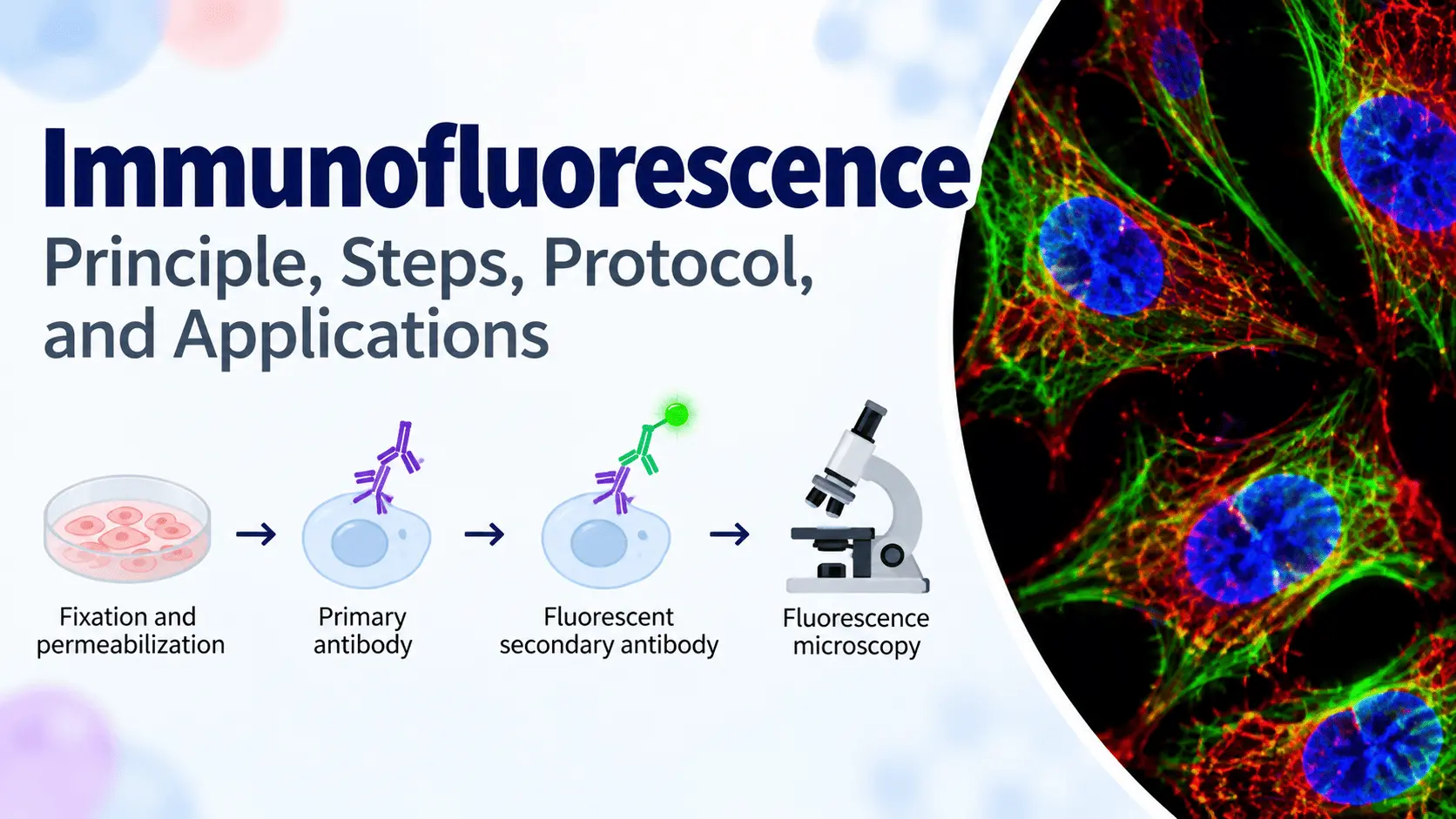
Immunofluorescence is a widely used laboratory technique for detecting and localizing specific proteins or other antigens in cells and tissues. The technique combines the specificity of antibody–antigen interactions with the ability of fluorescent molecules to produce visible signals under a fluorescence microscope.
Unlike techniques that simply measure whether a protein is present, immunofluorescence can provide important information about where a protein is located within a cell or tissue. Researchers can determine whether a target protein is found in the nucleus, cytoplasm, cell membrane, or specific cellular organelles.
Immunofluorescence is widely used in cell biology, immunology, microbiology, pathology, neuroscience, and cancer research. It can also be combined with confocal microscopy and image analysis to study protein localization and interactions in greater detail.
This guide explains the immunofluorescence principle, types, steps, applications, controls, troubleshooting, advantages, and limitations.
What Is Immunofluorescence?
Immunofluorescence is an antibody-based technique used to detect specific antigens in biological samples using fluorescently labeled antibodies.
The target antigen is first recognized by an antibody. A fluorescent molecule, known as a fluorophore, produces a detectable signal when exposed to light at an appropriate excitation wavelength.
The resulting fluorescence can be observed using a fluorescence microscope.
A typical immunofluorescence workflow can be summarized as:
Sample preparation → Fixation → Permeabilization → Blocking → Primary antibody → Washing → Secondary antibody → Counterstaining → Mounting → Fluorescence microscopy → Image analysis
One of the major advantages of immunofluorescence is that it allows researchers to visualize the spatial distribution of proteins within cells and tissues.
Immunofluorescence vs Immunohistochemistry
Compared with immunohistochemistry (IHC), both use antibodies to detect specific targets, but they differ mainly in their detection systems.
Immunofluorescence uses fluorescent labels to generate a signal, whereas immunohistochemistry commonly uses enzyme-based detection systems that produce a colored reaction product.
The technique is particularly useful when researchers need to visualize several targets simultaneously using different fluorophores.
IHC, on the other hand, is extensively used in diagnostic pathology and tissue-based biomarker analysis.
Types of Immunofluorescence
There are two major approaches to immunofluorescence:
- Direct immunofluorescence
- Indirect immunofluorescence
The main difference is where the fluorescent label is attached.
Direct Immunofluorescence
In direct immunofluorescence, the primary antibody is directly conjugated to a fluorophore.
The labeled primary antibody binds directly to the target antigen.
The workflow is relatively simple:
Target antigen → Fluorescently labeled primary antibody → Fluorescence signal
The main advantage is that fewer incubation steps are required.
However, direct immunofluorescence may provide lower signal amplification because each target molecule is generally detected by the labeled primary antibody.
Indirect Immunofluorescence
In indirect immunofluorescence, the primary antibody is not directly labeled.
Instead, a fluorescently labeled secondary antibody binds to the primary antibody.
The workflow is:
Target antigen → Primary antibody → Fluorescently labeled secondary antibody → Fluorescence signal
Indirect immunofluorescence is widely used because multiple secondary antibodies can potentially bind to a primary antibody, increasing the detectable signal.
It also provides greater flexibility because the same labeled secondary antibody can be used with different primary antibodies from the appropriate host species.
Immunofluorescence Principle
The immunofluorescence principle is based on specific antigen–antibody recognition combined with fluorescence detection.
The target antigen is present within a biological sample such as a cultured cell or tissue section. An antibody specifically recognizes this antigen.
The antibody is either directly conjugated to a fluorophore or detected using a fluorescent secondary antibody.
When the fluorophore is exposed to its appropriate excitation wavelength, it absorbs energy and subsequently emits light at a longer wavelength.
This emitted fluorescence is detected using a fluorescence microscope.
Antigen–Antibody Recognition
Antibody specificity is central to the immunofluorescence technique.
The primary antibody recognizes a specific epitope on the target antigen. The specificity of this interaction allows researchers to identify the location of the protein or other target within the sample.
However, antibody specificity is not absolute in every experimental context. Cross-reactivity and nonspecific binding can produce background staining or misleading signals.
For this reason, appropriate controls are essential.
Fluorescent Labels
Several fluorophores can be used for immunofluorescence.
Common examples include:
- FITC
- Alexa Fluor dyes
- Cy3
- Cy5
Different fluorophores have different excitation and emission characteristics.
Using fluorophores with distinct emission spectra allows researchers to perform multicolor immunofluorescence, in which several targets can be visualized in the same sample.
DAPI Nuclear Staining
DAPI (4′,6-diamidino-2-phenylindole) is commonly used as a nuclear counterstain.
It binds strongly to DNA and produces fluorescence that allows cell nuclei to be visualized.
This is particularly useful for determining the subcellular localization of a target protein relative to the nucleus.
For example, if a target protein is detected in the cytoplasm while DAPI identifies the nucleus, researchers can determine the approximate cellular distribution of the protein.
Fluorescence Microscopy
After staining, samples are examined using a fluorescence microscope.
The microscope uses appropriate filters or optical systems to excite the fluorophores and detect their emitted fluorescence.
Different fluorescence channels can be used to visualize different fluorophores.
For more advanced imaging, confocal microscopy can be used to obtain optical sections and improve visualization of fluorescent structures within thicker samples.
Immunofluorescence Steps and Protocol
A typical immunofluorescence protocol consists of several important stages. Exact conditions depend on the sample, target antigen, antibody, and imaging system.
1. Sample Preparation
Immunofluorescence can be performed using different types of biological samples, including:
- Cultured cells
- Tissue sections
- Frozen tissue
- Fixed tissue
- Biological specimens
For cultured cells, cells are commonly grown on coverslips or specialized imaging surfaces.
For tissue samples, appropriate sectioning and preparation are required before staining.
2. Fixation
Fixation preserves cellular structures and immobilizes proteins within the sample.
Common fixation approaches include chemical fixation using agents such as formaldehyde or paraformaldehyde.
The fixation conditions need to be optimized because excessive fixation can sometimes reduce antibody accessibility to target epitopes.
The choice of fixation method can therefore influence staining quality.
3. Permeabilization
Permeabilization is particularly important when the target protein is located inside the cell.
Permeabilizing agents create pores in cellular membranes, allowing antibodies to access intracellular targets.
This step may not be necessary for some surface or extracellular targets.
The appropriate permeabilization conditions depend on the biological sample and target protein.
4. Blocking
After fixation and, when necessary, permeabilization, the sample is treated with a blocking solution.
The purpose of blocking is to reduce nonspecific antibody binding.
Blocking reagents can occupy nonspecific binding sites, helping improve the signal-to-background ratio.
The optimal blocking reagent and conditions depend on the antibodies and sample type.
5. Primary Antibody Incubation
The sample is incubated with a primary antibody that specifically recognizes the target antigen.
Important factors include:
- Antibody specificity
- Antibody concentration
- Incubation time
- Incubation temperature
- Sample permeability
Following the manufacturer’s recommendations is a useful starting point, but experimental optimization may be necessary.
6. Washing
After primary antibody incubation, the sample is washed to remove unbound antibodies.
Effective washing is important for reducing nonspecific fluorescence.
Insufficient washing can contribute to high background, while overly harsh conditions may reduce specific signal.
7. Secondary Antibody Incubation
For indirect immunofluorescence, the sample is incubated with a fluorescently labeled secondary antibody.
The secondary antibody should recognize the host species of the primary antibody.
For example, if the primary antibody was produced in a rabbit, an appropriate anti-rabbit secondary antibody can be used.
For multicolor experiments, secondary antibodies with spectrally distinct fluorophores can be selected.
8. Nuclear Counterstaining
A nuclear stain such as DAPI can be added to visualize cell nuclei.
Counterstaining provides useful spatial information and makes it easier to interpret the localization of the target protein.
9. Mounting
After staining and washing, samples are mounted for microscopy.
Mounting media can help preserve the fluorescent signal and maintain the sample under the coverslip.
For fluorescent imaging, antifade mounting media may be used to reduce photobleaching.
10. Fluorescence Microscopy and Image Acquisition
The stained sample is examined under a fluorescence microscope.
The appropriate imaging channel is selected according to the fluorophore being used.
During image acquisition, researchers should avoid excessive exposure because intense illumination can cause photobleaching and may produce saturated signals.
For multicolor experiments, individual channels can be recorded separately and subsequently combined into a composite image.
11. Image Analysis
The resulting images can be analyzed qualitatively or quantitatively.
Depending on the research question, researchers may measure:
- Fluorescence intensity
- Number of positive cells
- Protein localization
- Colocalization
- Percentage of stained area
- Changes in fluorescence between experimental groups
Image analysis should use consistent acquisition settings and appropriate controls whenever quantitative comparisons are made.
Immunofluorescence Applications in Biology and Medicine
Immunofluorescence has applications across many areas of biological and biomedical research.
Protein Localization
One of the most important applications is determining the localization of a protein within a cell.
Researchers can determine whether a protein is predominantly:
- Nuclear
- Cytoplasmic
- Membrane-associated
- Mitochondrial
- Golgi-associated
- Endoplasmic-reticulum-associated
This spatial information can provide clues about the biological function of the protein.
Cell Biology
Immunofluorescence is extensively used in cell biology to visualize cellular structures and proteins.
For example, antibodies can be used to investigate:
- Cytoskeletal proteins
- Cell-cycle proteins
- Membrane proteins
- Organelle-associated proteins
- Signaling molecules
- Cell adhesion proteins
Immunofluorescence can therefore help researchers understand how proteins are distributed within cells and how their localization changes under different conditions.
Immunology
In immunology, immunofluorescence can be used to identify immune-cell markers and investigate the distribution of immune-related proteins.
Researchers can examine proteins associated with:
- T cells
- B cells
- Macrophages
- Dendritic cells
- Neutrophils
- Other immune-cell populations
Multicolor immunofluorescence can be particularly useful when several cellular markers need to be examined simultaneously.
Immunofluorescence in Cancer Research
Immunofluorescence is an important technique in cancer biology research because cancer development involves changes in protein expression, localization, signaling, and cellular organization.
Researchers can use immunofluorescence to investigate:
- Tumor-associated proteins
- Oncogenic signaling pathways
- Tumor suppressor proteins
- Cell proliferation
- Apoptosis
- Epithelial–mesenchymal transition
- Cancer cell phenotypes
- Tumor microenvironment components
- Immune-cell populations within tumors
For example, researchers can use different fluorescent antibodies to identify cancer cells and immune-cell populations within the same tissue.
Immunofluorescence can also be used to investigate the localization of signaling proteins in cancer cells and determine whether experimental treatments alter their intracellular distribution.
Histopathology and Disease Research
Immunofluorescence can also be used to study disease-associated proteins in tissue samples.
Applications include research into:
- Neurological diseases
- Autoimmune diseases
- Infectious diseases
- Kidney diseases
- Cancer
- Inflammatory diseases
By combining antibody specificity with spatial visualization, researchers can investigate the distribution of disease-associated molecules within tissues.
Immunofluorescence Controls, Troubleshooting, and Limitations
Proper controls and experimental optimization are essential for obtaining reliable immunofluorescence results.
Essential Immunofluorescence Controls
Several controls can help distinguish specific staining from background fluorescence.
Negative Control
A negative control is expected to produce little or no specific fluorescence.
It can help determine whether the observed signal is associated with nonspecific staining.
No-Primary-Antibody Control
In this control, the primary antibody is omitted while other staining steps are maintained.
If substantial fluorescence remains, it may indicate nonspecific binding of the secondary antibody or autofluorescence.
Positive Control
A positive control contains a sample known to express the target antigen.
This helps verify that the antibody and staining procedure are capable of detecting the target.
Isotype Control
An isotype control uses an antibody of the same immunoglobulin class as the primary antibody but without specificity for the target antigen.
Isotype controls can sometimes help assess nonspecific antibody interactions, although their usefulness depends on the experimental design.
Single-Color Controls
For multicolor immunofluorescence, single-color controls can help determine whether fluorescence from one channel is detected in another channel.
These controls are particularly useful when fluorophores have overlapping emission spectra.
Common Immunofluorescence Problems
High Background
High background fluorescence can make specific staining difficult to distinguish.
Potential causes include:
- Insufficient blocking
- Excessive antibody concentration
- Inadequate washing
- Nonspecific antibody binding
- Autofluorescence
- Excessive exposure during imaging
Possible solutions include optimizing antibody concentrations, improving washing, adjusting blocking conditions, and reducing imaging exposure.
Weak Fluorescence Signal
Weak fluorescence may result from:
- Low target abundance
- Poor antibody affinity
- Incorrect antibody concentration
- Inefficient permeabilization
- Inappropriate fixation
- Photobleaching
Checking the antibody and optimizing sample preparation can help improve the signal.
Nonspecific Staining
Nonspecific staining can occur when antibodies bind to unintended targets.
Possible causes include antibody cross-reactivity, inadequate blocking, or excessive antibody concentration.
Using validated antibodies and appropriate controls is important for minimizing this problem.
Uneven Staining
Uneven fluorescence across the sample may result from inadequate sample preparation, inconsistent antibody penetration, or incomplete washing.
For tissue samples, sample thickness and accessibility of the target antigen can also influence staining.
Photobleaching
Photobleaching occurs when fluorescent molecules lose their ability to emit fluorescence after prolonged exposure to excitation light.
To reduce photobleaching:
- Minimize unnecessary exposure to light.
- Use appropriate imaging settings.
- Avoid repeated imaging when possible.
- Use suitable antifade mounting media.
- Store fluorescent samples appropriately.
Advantages of Immunofluorescence
Immunofluorescence offers several important advantages:
- High specificity through antibody-based detection.
- Visualization of protein localization.
- Ability to analyze cells and tissues spatially.
- Possibility of multicolor staining.
- Compatibility with fluorescence and confocal microscopy.
- Ability to investigate several cellular markers simultaneously.
- Useful for both qualitative and quantitative imaging studies.
Limitations of Immunofluorescence
Despite its advantages, immunofluorescence has several limitations.
These include:
- Photobleaching
- Autofluorescence
- Nonspecific antibody binding
- Dependence on antibody quality
- Potential spectral overlap between fluorophores
- Need for fluorescence microscopy equipment
- Time-consuming optimization
- Difficulty interpreting fluorescence intensity as a direct measure of protein abundance
Importantly, stronger fluorescence does not necessarily mean that a sample contains proportionally more protein. Experimental conditions, antibody affinity, fluorophore properties, imaging settings, and sample preparation can all influence fluorescence intensity.
Conclusion
Immunofluorescence is a powerful biology technique for detecting and localizing specific proteins and antigens in cells and tissues. By combining antigen–antibody recognition with fluorescent detection, the technique provides spatial information that is extremely valuable for understanding cellular organization and biological processes.
The main stages of immunofluorescence include sample preparation, fixation, permeabilization, blocking, antibody incubation, washing, counterstaining, mounting, fluorescence microscopy, and image analysis.
Both direct and indirect immunofluorescence approaches can be used, with indirect immunofluorescence offering greater flexibility and potential signal amplification.
The technique has broad applications in cell biology, immunology, pathology, and cancer research, including protein localization, immune-cell characterization, tumor biology, and investigation of disease-associated biomarkers.
Reliable immunofluorescence results depend on appropriate antibody selection, optimized staining conditions, suitable controls, and careful image acquisition. When these factors are properly controlled, immunofluorescence provides a powerful way to visualize molecular processes directly within their cellular and tissue context.


