

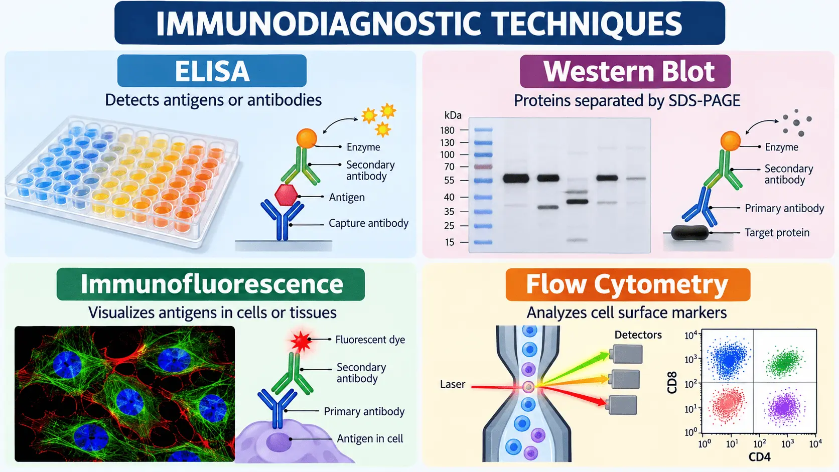
Immunodiagnostic techniques are laboratory methods that use the specificity of antigen–antibody interactions to detect, identify, or measure biological molecules and cellular markers. These techniques are widely used in clinical diagnosis, biomedical research, immunology, microbiology, and cancer biology.
By exploiting the ability of antibodies to recognize specific antigens, immunodiagnostic methods can detect pathogens, measure antibodies in patient samples, identify disease-associated proteins, and characterize specific cell populations.
Among the most commonly used immunodiagnostic techniques are ELISA, Western blotting, immunofluorescence, and flow cytometry. Although all four methods rely on antibody-based detection, they differ in their principles, sample types, detection systems, and applications.
1. ELISA
Enzyme-linked immunosorbent assay (ELISA) is one of the most widely used immunodiagnostic techniques for detecting and quantifying antigens or antibodies in biological samples.
ELISA is commonly performed using microtiter plates in which antigens or antibodies are immobilized on a solid surface. An enzyme-linked antibody is then used to generate a measurable signal when an appropriate substrate is added.
Principle of ELISA
The fundamental principle of ELISA is based on the highly specific interaction between an antigen and its corresponding antibody.
Depending on the type of ELISA, the target molecule may be captured directly or indirectly. After the antigen–antibody reaction occurs, an enzyme-linked detection system produces a colorimetric, fluorescent, or chemiluminescent signal.
The intensity of the signal can be related to the amount of target molecule present in the sample.
Main types of ELISA
Several ELISA formats are commonly used:
Direct ELISA:
An enzyme-labeled primary antibody directly recognizes the target antigen.
Indirect ELISA:
An unlabeled primary antibody binds the antigen and is detected by an enzyme-labeled secondary antibody.
Sandwich ELISA:
The target antigen is captured between two antibodies. This format is particularly useful for detecting proteins present at relatively low concentrations.
Competitive ELISA:
The sample antigen competes with a labeled antigen or another molecule for antibody binding. This format can be useful when the target molecule is small or has only one available antibody-binding site.
Applications of ELISA
ELISA has numerous applications in clinical and research laboratories. It can be used to detect:
- Antibodies produced during infections
- Microbial antigens
- Hormones
- Cytokines
- Tumor-associated biomarkers
- Allergens
- Autoantibodies
- Other proteins and biological molecules
For example, antibody-based ELISA assays can be used to determine whether an individual has developed an immune response to a particular infectious agent.
Advantages and limitations
ELISA is relatively simple, scalable, and suitable for processing large numbers of samples. Its quantitative nature also makes it useful for measuring concentrations of specific targets.
However, ELISA depends strongly on antibody specificity and assay design. Cross-reactivity can produce false-positive results, while insufficient sensitivity can lead to false-negative results.
2. Western Blot
Western blotting, also called immunoblotting, is an immunological technique used to detect a specific protein within a complex mixture of proteins.
Unlike ELISA, which commonly measures an antigen or antibody directly in a sample, Western blot combines protein separation by SDS-PAGE with antibody-based detection.
Principle of Western blot
The first step is the extraction of proteins from cells, tissues, or other biological samples.
The proteins are then separated according to their molecular weight using sodium dodecyl sulfate–polyacrylamide gel electrophoresis (SDS-PAGE).
After electrophoresis, the separated proteins are transferred from the gel onto a membrane, commonly a PVDF or nitrocellulose membrane.
The membrane is then incubated with a primary antibody that specifically recognizes the protein of interest. A labeled secondary antibody may subsequently bind to the primary antibody.
Finally, the target protein is visualized using an appropriate detection system.
Major steps of Western blotting
The general workflow includes:
- Protein extraction
- Protein quantification
- Sample preparation
- SDS-PAGE separation
- Transfer to a membrane
- Blocking
- Primary antibody incubation
- Secondary antibody incubation
- Signal detection
- Protein band analysis
The position of the detected band provides information about the approximate molecular weight of the protein, while band intensity can be used for relative quantification under appropriate experimental conditions.
Applications of Western blot
Western blotting is widely used to investigate protein expression and signaling pathways.
Applications include:
- Detection of specific proteins
- Analysis of protein expression
- Verification of antibodies
- Investigation of signaling pathways
- Detection of post-translationally modified proteins
- Confirmation of findings from other protein assays
- Biomedical and cancer research
In cancer biology, for example, Western blotting can be used to investigate changes in proteins involved in cell proliferation, apoptosis, migration, invasion, and signal transduction.
Advantages and limitations
A major advantage of Western blotting is that it combines antibody specificity with protein separation. This allows researchers to distinguish a target protein from other proteins in a complex sample based partly on molecular size.
However, Western blotting is relatively labor-intensive and requires several experimental steps. Results can also be affected by antibody specificity, protein transfer efficiency, loading differences, and detection conditions.
3. Immunofluorescence
Immunofluorescence (IF) is an antibody-based technique used to visualize specific antigens in cells or tissues.
Unlike ELISA and Western blotting, which generally provide quantitative or semi-quantitative measurements, immunofluorescence provides important spatial information about where a target protein is located.
Fluorescently labeled antibodies are used to identify the target antigen, and the resulting fluorescence can be observed using a fluorescence or confocal microscope.
Principle of immunofluorescence
The basic principle involves using antibodies that specifically recognize an antigen within a biological sample.
There are two major approaches.
Direct immunofluorescence uses a fluorescently labeled primary antibody that binds directly to the target antigen.
Indirect immunofluorescence uses an unlabeled primary antibody followed by a fluorescently labeled secondary antibody.
Indirect immunofluorescence can provide signal amplification because several secondary antibodies may bind to a primary antibody.
Immunofluorescence workflow
A typical immunofluorescence experiment involves:
- Preparation of cells or tissue sections
- Fixation
- Permeabilization when intracellular targets are studied
- Blocking of nonspecific binding
- Primary antibody incubation
- Fluorescent secondary antibody incubation, when applicable
- Nuclear or cellular counterstaining
- Microscopic imaging
- Image analysis
Fluorescent dyes such as fluorophores attached to antibodies emit light at specific wavelengths when excited by an appropriate light source.
Applications of immunofluorescence
Immunofluorescence is particularly useful for determining the cellular and subcellular localization of proteins.
It can be used to investigate:
- Nuclear proteins
- Membrane proteins
- Cytoplasmic proteins
- Cell-surface markers
- Tissue-specific antigens
- Protein localization
- Cellular signaling
- Disease-associated biomarkers
In cancer research, immunofluorescence can help determine whether a protein is localized to the nucleus, cytoplasm, cell membrane, or other cellular compartments.
Advantages and limitations
The major advantage of immunofluorescence is its ability to provide visual information about antigen distribution and localization.
Multiple fluorescent markers can also be combined to visualize different targets in the same sample.
However, immunofluorescence requires appropriate microscopy and careful optimization. Background fluorescence, nonspecific antibody binding, photobleaching, and antibody cross-reactivity can complicate interpretation.
4. Flow Cytometry
Flow cytometry is a powerful immunodiagnostic and cell-analysis technique that allows individual cells to be analyzed rapidly as they pass through a laser beam.
It is particularly useful for identifying and quantifying cell populations according to their surface or intracellular markers.
Antibodies conjugated to fluorescent molecules are commonly used to identify specific proteins on or inside cells.
Principle of flow cytometry
In flow cytometry, cells are suspended in a liquid and passed individually through an interrogation point where they interact with one or more laser beams.
The instrument measures different properties of each cell.
These commonly include:
- Forward scatter
- Side scatter
- Fluorescence intensity
Forward and side scatter provide information related to cellular characteristics such as size and internal complexity, while fluorescent signals indicate the presence and relative abundance of specific markers.
Immunophenotyping
One of the most important applications of flow cytometry is immunophenotyping.
In immunophenotyping, fluorescently labeled antibodies recognize specific cell-surface or intracellular molecules.
For example, different antibodies can be used to distinguish populations of immune cells according to their characteristic markers.
Multiple fluorophores can be used simultaneously, allowing several markers to be analyzed on the same cell.
This makes flow cytometry particularly useful when researchers need to determine not only whether a marker is present but also which cells express it.
Applications of flow cytometry
Flow cytometry has applications in:
- Immunophenotyping
- Hematological disease investigation
- Leukemia and lymphoma characterization
- Immune-cell analysis
- Cell-cycle analysis
- Apoptosis studies
- Stem-cell research
- Cancer research
- Measurement of intracellular proteins
- Quantification of cell populations
In cancer biology, flow cytometry can be used to characterize tumor cells and immune-cell populations within a tumor or biological sample.
Advantages and limitations
Flow cytometry can analyze thousands of cells rapidly and can distinguish multiple cell populations within a heterogeneous sample.
Its ability to combine several fluorescent markers also makes it highly informative for complex cellular analyses.
However, flow cytometry requires specialized instrumentation and expertise in experimental design, compensation, gating, and data interpretation. Poor antibody selection or inappropriate gating strategies can lead to misleading results.
Comparing the Four Immunodiagnostic Techniques
Although ELISA, Western blot, immunofluorescence, and flow cytometry all use antigen–antibody recognition, they answer different biological questions.
| Technique | Main target | Main purpose | Typical output |
|---|---|---|---|
| ELISA | Antigens or antibodies | Detection and quantification | Color/fluorescence/chemiluminescence signal |
| Western blot | Specific proteins | Protein detection and analysis | Protein bands |
| Immunofluorescence | Cellular or tissue antigens | Localization of proteins | Fluorescence images |
| Flow cytometry | Cell-surface or intracellular markers | Cell identification and quantification | Fluorescence and cell-population data |
The choice of technique therefore depends on the biological question.
If the objective is to measure an antigen or antibody in a sample, ELISA may be appropriate.
When the objective is to detect a particular protein after separating proteins by molecular weight, Western blotting can be used.
If researchers need to determine where a protein is located inside cells or tissues, immunofluorescence provides spatial information.
If the goal is to identify and quantify different cell populations based on molecular markers, flow cytometry is particularly useful.
Conclusion
Immunodiagnostic techniques are essential tools in modern biomedical science because they exploit the specificity of antigen–antibody interactions to detect molecules and characterize cells.
ELISA is widely used for antigen and antibody detection and quantification. Western blotting combines protein separation with antibody-based detection to identify specific proteins. Immunofluorescence allows researchers to visualize antigens within cells and tissues, providing valuable spatial information. Flow cytometry enables rapid multiparametric analysis of individual cells based on surface and intracellular markers.
Together, these techniques support applications ranging from infectious disease diagnosis and immunology to cancer research, biomarker investigation, and cellular characterization. Understanding the principle and appropriate application of each method is essential for selecting the right immunodiagnostic approach for a particular research or clinical question.


