

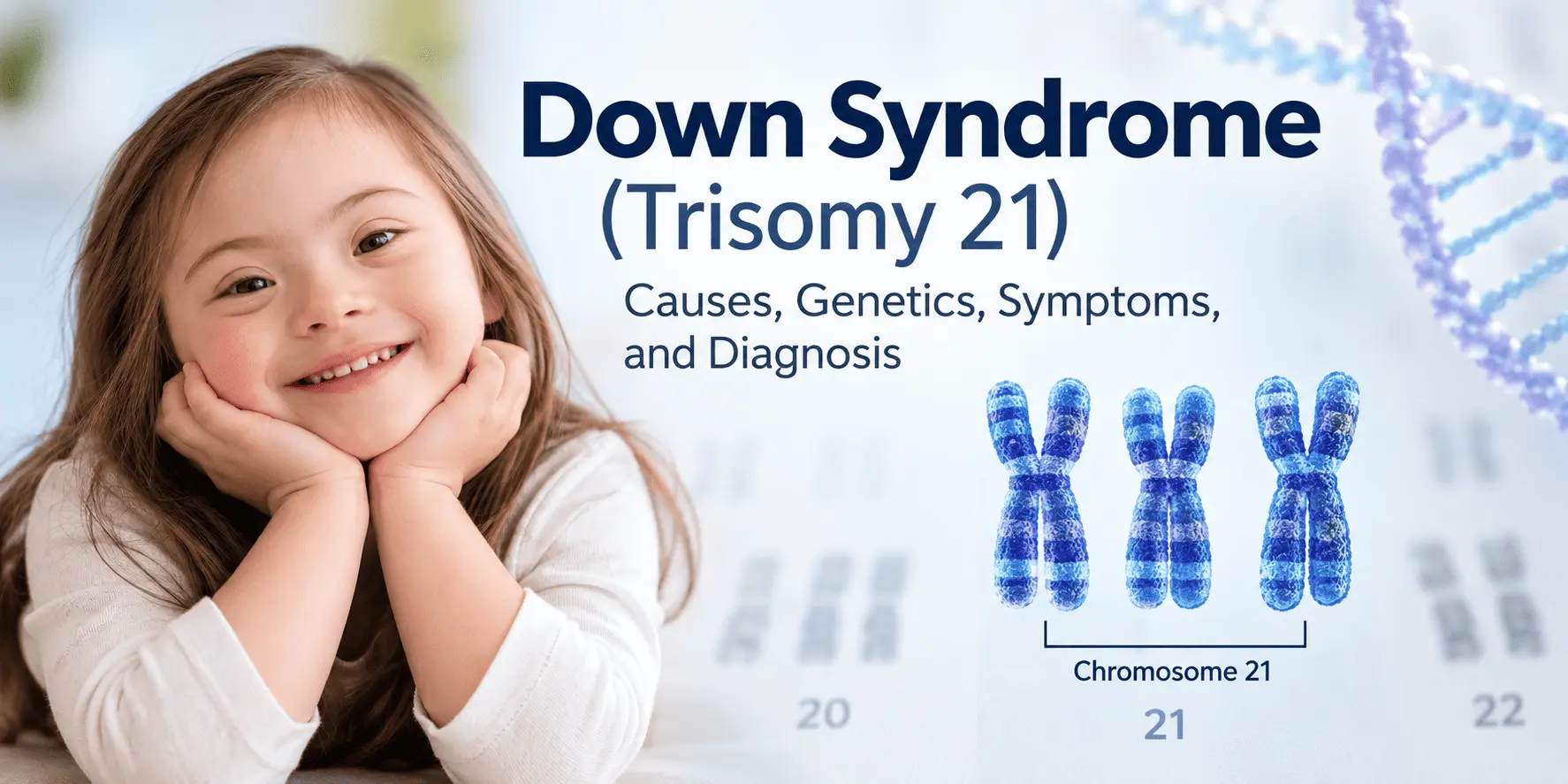
Down syndrome, also known as trisomy 21, is a genetic condition caused by the presence of extra genetic material from chromosome 21. It is the most common chromosomal condition associated with intellectual and developmental differences and can also affect physical development and overall health.
Down syndrome occurs because cells contain an additional copy of chromosome 21 or additional chromosome 21 material. The extra genetic material changes the expression of genes involved in development, resulting in characteristic physical features, developmental differences, and an increased risk of certain medical conditions.
From a genetics perspective, Down syndrome is particularly important because it illustrates how a change in chromosome number, known as aneuploidy, can influence human development.
What Is Down Syndrome (Trisomy 21)?
Humans normally have 46 chromosomes arranged in 23 pairs. Each pair contains one chromosome inherited from the mother and one from the father. Chromosome 21 is one of the smallest human chromosomes but contains many genes involved in biological processes important for development.
In most cases of Down syndrome, a person has three copies of chromosome 21 instead of two. This is called trisomy 21.
The additional chromosome 21 increases the amount of genetic material present in cells. This phenomenon is often described as a gene dosage effect: having an extra copy of a chromosome can increase the expression of some genes and disrupt normal biological pathways.
Down syndrome is not caused by a single gene mutation. Instead, it is a chromosomal disorder involving many genes located on chromosome 21.
There are three major genetic forms of Down syndrome:
- Trisomy 21: An extra complete chromosome 21 is present in the cells.
- Translocation Down syndrome: Extra chromosome 21 material is attached to another chromosome.
- Mosaic Down syndrome: Only some cells contain the extra chromosome 21, while other cells have the usual chromosome number.
Although these forms have the same general genetic consequence—extra chromosome 21 material—the mechanism by which the chromosomal abnormality arises differs.
Full Trisomy 21
Full trisomy 21 accounts for the large majority of Down syndrome cases. In this form, essentially all cells contain three copies of chromosome 21.
The extra chromosome usually results from an error during the formation of an egg or sperm. This error is called nondisjunction.
Translocation Down Syndrome
In translocation Down syndrome, an extra copy or portion of chromosome 21 becomes attached to another chromosome.
A common form involves a Robertsonian translocation, in which chromosome 21 material becomes attached to another acrocentric chromosome, frequently chromosome 14 or another chromosome 21.
Unlike most cases of full trisomy 21, translocation Down syndrome can sometimes be associated with a balanced translocation in a parent. A parent carrying a balanced translocation may have no clinical features because there is no significant loss or gain of genetic material. However, chromosome rearrangement during reproduction can result in an embryo receiving extra chromosome 21 material.
For this reason, identifying translocation Down syndrome can be particularly important for genetic counseling.
Mosaic Down Syndrome
Mosaic Down syndrome occurs when an individual has two or more genetically different cell populations.
Some cells have the usual chromosome complement of 46 chromosomes, while other cells contain an additional chromosome 21.
Mosaicism generally develops because of a chromosome-separation error occurring after fertilization during early cell division. The proportion and distribution of cells carrying the extra chromosome can vary considerably between individuals.
Because of this variation, the characteristics associated with mosaic Down syndrome can also vary.
Genetic Causes and Mechanisms of Down Syndrome
The fundamental genetic cause of Down syndrome is the presence of extra chromosome 21 material. However, the cellular mechanisms that produce this abnormality can differ.
Chromosomal Nondisjunction
The most common mechanism is meiotic nondisjunction.
During meiosis, chromosome pairs normally separate so that each egg or sperm receives one chromosome from each pair. After fertilization, the resulting embryo normally receives two copies of each autosomal chromosome—one from each parent.
If chromosome 21 fails to separate correctly during meiosis, a reproductive cell can receive two copies of chromosome 21 instead of one.
If that cell participates in fertilization, the resulting embryo can have three copies of chromosome 21.
In simplified terms:
Normal:
Egg → 1 chromosome 21
Sperm → 1 chromosome 21
Embryo → 2 chromosome 21 copies
Nondisjunction:
Egg or sperm → 2 chromosome 21 copies
Other reproductive cell → 1 chromosome 21 copy
Embryo → 3 chromosome 21 copies
Nondisjunction can occur during the formation of either the egg or sperm, although chromosome 21 nondisjunction in the maternal germline accounts for most cases of full trisomy 21.
Maternal Age and Nondisjunction
The probability of having a pregnancy affected by trisomy 21 increases with maternal age, particularly as maternal age advances.
One important biological explanation involves age-related changes in oocytes. Human oocytes begin meiosis before birth and remain arrested for many years before completing the process around ovulation and fertilization. The mechanisms responsible for maintaining accurate chromosome segregation can become less reliable with increasing maternal age.
However, it is important to understand that Down syndrome can occur in pregnancies at any maternal age. Because younger women have more pregnancies overall, a substantial number of babies with Down syndrome are also born to younger mothers.
Maternal age is therefore a risk factor, not a direct cause of Down syndrome.
Robertsonian Translocation
A smaller proportion of Down syndrome cases result from chromosome translocation.
A Robertsonian translocation occurs when the long arms of two acrocentric chromosomes become joined. Chromosomes 13, 14, 15, 21, and 22 are the human acrocentric chromosomes.
When chromosome 21 material is involved in a Robertsonian translocation, an individual may have extra chromosome 21 genetic material despite not having three separate chromosome 21 molecules.
Some translocation cases occur as new chromosome rearrangements, while others are inherited from a parent carrying a balanced translocation.
This is why chromosome analysis can be important after a diagnosis of Down syndrome, particularly when determining the possibility of recurrence in future pregnancies.
Mosaicism
Mosaic Down syndrome develops through a different mechanism.
Instead of the chromosome abnormality being present from the formation of the reproductive cell, a chromosome-separation error can occur during an early mitotic cell division after fertilization.
As a result, two populations of cells may develop:
- Cells with the usual chromosome number
- Cells containing an additional chromosome 21
The proportion of trisomic cells can differ between tissues, which contributes to variation in the characteristics of mosaic Down syndrome.
Gene Dosage Effects
Why does an extra chromosome cause developmental and physiological changes?
One major concept is gene dosage.
An additional chromosome 21 means that many genes located on chromosome 21 are present in three copies rather than two. Increased gene dosage can alter the production of proteins and influence cellular pathways.
Researchers have identified several chromosome 21 genes that may contribute to specific features associated with Down syndrome. Examples include DYRK1A, RCAN1, and APP, among others.
However, Down syndrome cannot be explained by one specific gene. It results from the combined effects of increased dosage of numerous genes and interactions between genetic and environmental factors.
Signs, Symptoms, and Health Characteristics of Down Syndrome
Down syndrome can influence multiple aspects of development and health. However, there is considerable variation between individuals.
Not every person with Down syndrome has the same physical characteristics, developmental profile, or medical conditions.
Physical Characteristics
Common physical characteristics may include:
- Low muscle tone, known as hypotonia
- Short stature
- A relatively flat facial profile
- Upward-slanting palpebral fissures
- A shorter neck
- Small ears
- A single transverse palmar crease in some individuals
- Increased joint flexibility
These characteristics vary in their presence and degree.
Physical features alone are not sufficient to establish a definitive diagnosis. Chromosome analysis or another appropriate genetic test is used to confirm the chromosomal abnormality.
Development and Learning
People with Down syndrome commonly experience some degree of intellectual disability and developmental delay.
Developmental milestones such as sitting, walking, speaking, and acquiring certain cognitive skills may occur later than in children without the condition.
Language development can be particularly variable. Many individuals develop strong social communication skills but may experience difficulties with expressive language and speech.
Early developmental support, education, speech therapy, occupational therapy, and physical therapy can help individuals develop skills and participate more independently in daily life.
Congenital Heart Conditions
Congenital heart defects are relatively common in people with Down syndrome.
Examples include:
- Atrioventricular septal defects
- Ventricular septal defects
- Atrial septal defects
- Other structural abnormalities of the heart
Because of this increased risk, cardiac evaluation is an important component of medical care following a diagnosis.
Hearing and Vision Problems
Hearing problems can occur more frequently in people with Down syndrome. Causes can include recurrent ear infections, fluid accumulation in the middle ear, and sensorineural hearing impairment.
Vision problems may include refractive errors, strabismus, cataracts, or other eye conditions.
Regular hearing and ophthalmologic assessments can help identify problems early.
Thyroid Disorders
Thyroid dysfunction, particularly hypothyroidism, occurs more frequently in people with Down syndrome than in the general population.
Regular thyroid screening is therefore commonly incorporated into medical follow-up.
Gastrointestinal Conditions
Some individuals with Down syndrome have gastrointestinal abnormalities, including congenital conditions such as duodenal atresia and Hirschsprung disease.
Other gastrointestinal problems can also occur and may require medical evaluation and treatment.
Sleep-Related Breathing Disorders
Obstructive sleep apnea is relatively common in people with Down syndrome.
Anatomical characteristics, muscle tone, and airway structure can contribute to airway obstruction during sleep.
Symptoms such as loud snoring, pauses in breathing, restless sleep, or excessive daytime sleepiness may warrant medical evaluation.
How Is Down Syndrome Diagnosed?
Down syndrome can be identified through prenatal screening, prenatal diagnostic testing, or chromosome testing after birth.
It is important to distinguish between screening tests and diagnostic tests.
A screening test estimates the likelihood that a fetus has a chromosomal condition. A diagnostic test can establish whether the chromosomal abnormality is actually present.
Prenatal Screening
Several screening approaches can estimate the likelihood of trisomy 21.
Ultrasound examination can identify certain fetal features associated with chromosomal abnormalities. For example, measurement of nuchal translucency during the first trimester can contribute to risk assessment.
Maternal blood tests can also measure biochemical markers associated with chromosomal abnormalities.
Another important screening method is cell-free DNA testing, sometimes called noninvasive prenatal testing (NIPT). This test analyzes DNA fragments circulating in the pregnant person’s blood and can provide a highly effective screening assessment for trisomy 21.
However, cell-free DNA testing remains a screening test, not a definitive diagnostic test.
Chorionic Villus Sampling
Chorionic villus sampling (CVS) is a prenatal diagnostic procedure that obtains a sample of placental tissue.
The fetal genetic material in the sample can then be analyzed for chromosomal abnormalities, including trisomy 21.
CVS is generally performed earlier in pregnancy than amniocentesis.
Amniocentesis
Amniocentesis involves collecting a small amount of amniotic fluid containing fetal cells.
These cells can be analyzed to determine the fetal chromosome complement.
Both CVS and amniocentesis are invasive diagnostic procedures and carry procedure-related risks, so decisions about prenatal testing should be discussed with an appropriately qualified healthcare professional.
Diagnosis After Birth
After birth, a healthcare professional may suspect Down syndrome based on physical characteristics and clinical findings.
However, clinical examination alone does not establish the chromosomal diagnosis.
A blood sample can be analyzed using chromosome analysis, most commonly through karyotyping.
Karyotyping
Karyotyping allows chromosomes to be visualized and organized according to their size and structural characteristics.
In a typical case of full trisomy 21, the karyotype demonstrates three copies of chromosome 21.
For example, instead of the usual chromosome number:
46,XX or 46,XY
a person with full trisomy 21 may have:
47,XX,+21 or 47,XY,+21
The notation indicates an additional chromosome 21.
Karyotyping can also help distinguish full trisomy 21 from translocation Down syndrome.
Other laboratory techniques, such as fluorescence in situ hybridization (FISH) or chromosomal microarray testing, may also be used in specific diagnostic contexts. The appropriate test depends on the clinical situation and the question being investigated.
Down Syndrome Risk Factors, Genetics, and Management
Several factors are associated with an increased probability of Down syndrome, although none alone guarantees that a pregnancy will be affected.
Maternal Age
Advanced maternal age is the most well-established population-level risk factor for a pregnancy affected by trisomy 21.
The risk increases as maternal age increases, particularly after age 35. However, Down syndrome can occur at any reproductive age.
Therefore, maternal age should be understood as a statistical risk factor rather than a direct cause.
Previous Pregnancy With Down Syndrome
A previous pregnancy affected by Down syndrome can increase the chance of Down syndrome occurring in a subsequent pregnancy.
The exact recurrence risk depends partly on the chromosomal mechanism involved in the previous pregnancy.
For example, recurrence considerations differ between:
- Full trisomy 21
- Translocation Down syndrome
- Mosaic Down syndrome
This makes determining the specific chromosome abnormality important when counseling families.
Parental Chromosomal Translocation
If Down syndrome results from a translocation, chromosome analysis of the parents may be recommended.
A parent carrying a balanced Robertsonian translocation involving chromosome 21 can have an increased chance of producing reproductive cells with an unbalanced chromosome complement.
Genetic counseling can help explain the specific chromosome rearrangement and its implications for future pregnancies.
Genetic Counseling
Counseling can be useful for families following a diagnosis of Down syndrome.
A genetic counselor can explain:
- The chromosome abnormality
- The mechanism responsible for it
- Whether the condition was likely inherited
- Recurrence risk
- Available prenatal testing options
- Implications for other family members when relevant
The genetic mechanism is particularly important when translocation Down syndrome is identified.
Medical Management
There is no single treatment that removes the extra chromosome 21. Instead, medical care focuses on monitoring health, treating associated conditions, and supporting development.
Depending on individual needs, care may involve:
- Pediatric or primary medical care
- Cardiology
- Audiology
- Ophthalmology
- Endocrinology
- Speech and language therapy
- Physical therapy
- Occupational therapy
- Developmental and educational support
Early Intervention
Early intervention programs can support children with Down syndrome in developing communication, motor, cognitive, and social skills.
Physical therapy can help address motor development and muscle tone. Speech and language therapy can support communication. Occupational therapy can help with fine motor abilities and everyday activities.
Educational approaches should be individualized according to the person’s developmental profile and needs.
Long-Term Health
People with Down syndrome can live into adulthood and participate in education, employment, relationships, and community life.
Health needs can change throughout life, making appropriate medical follow-up important.
Some medical conditions occur more frequently in adults with Down syndrome, so healthcare providers may recommend age-appropriate screening and monitoring.
The health and developmental profile of Down syndrome is highly variable, and the needs of one individual cannot be assumed to represent those of another.
Conclusion
Down syndrome (trisomy 21) is a chromosomal condition caused by the presence of extra genetic material from chromosome 21. The most common form results from meiotic nondisjunction and produces a complete extra chromosome 21.
Other cases result from chromosomal translocation or mosaicism. These mechanisms are important because they can influence the interpretation of genetic testing and, particularly in translocation cases, the recurrence risk for future pregnancies.
The extra chromosome 21 affects gene dosage and developmental processes, contributing to characteristic physical features, developmental differences, and an increased likelihood of certain medical conditions.
Diagnosis can involve prenatal screening, prenatal diagnostic procedures, and chromosome analysis after birth. Karyotyping remains particularly useful for determining the chromosomal basis of a confirmed diagnosis.
Understanding the genetics of Down syndrome provides an important foundation for diagnosis, genetic counseling, medical monitoring, early intervention, and individualized support.


, including its genetic causes, chromosome 21 abnormalities, symptoms, risk factors, diagnosis, and management.)