Cancer is one of the most significant challenges in modern medicine, affecting millions worldwide. Despite advancements, finding effective treatments remains a priority. Researchers are constantly exploring innovative approaches to better understand and combat this disease.
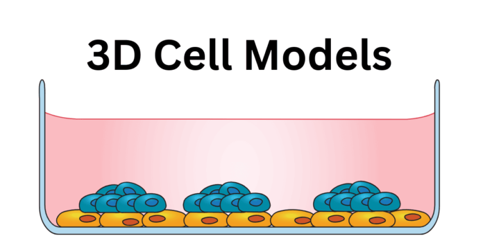
One such approach is the use of 3D cell models. Unlike traditional 2D cell cultures, 3D cell models offer a more accurate representation of the human body’s cellular environment. These models mimic the structure and function of actual tumor tissues, providing a powerful tool for studying cancer.
3D cell models are crucial in cancer research because they replicate the tumor microenvironment, allowing scientists to observe interactions between cancer cells and surrounding tissues, test treatments, and gain insights into cancer progression.
This blog post explores the future of cancer research through the lens of 3D cell models, highlighting their importance and potential in transforming cancer therapy.
Understanding 3D Cell Models
What are 3D Cell Models?
Definition and Basic Concept
3D cell models are advanced in vitro systems that replicate the three-dimensional architecture of human tissues. These models are composed of cells that grow in all directions, forming structures that closely resemble the natural organization of tissues within the body.
By providing a more accurate representation of the in vivo environment, 3D cell models enable researchers to study cellular behaviors and interactions in a setting that mimics real-life conditions.
Difference Between 2D and 3D Cell Cultures
Traditional 2D cell cultures involve growing cells on flat, rigid surfaces such as petri dishes or flasks. While these cultures have been instrumental in many scientific discoveries, they have significant limitations.
In 2D cultures, cells grow in a monolayer, which can alter their morphology, function, and interactions, making it difficult to accurately predict how cells behave in the body.
In contrast, 3D cell cultures allow cells to grow in a three-dimensional matrix, providing a more physiologically relevant environment. This allows cells to maintain their natural shape, function, and interactions with neighboring cells. Key differences between 2D and 3D cell cultures include:
- Cell Morphology: In 3D cultures, cells exhibit natural shapes and structures, while in 2D cultures, they often become flattened and stretched.
- Cell-Cell and Cell-Matrix Interactions: 3D models facilitate complex interactions between cells and their surrounding matrix, closely mimicking in vivo conditions. In 2D cultures, these interactions are limited and not representative of true tissue environments.
- Nutrient and Oxygen Gradients: 3D cultures can create gradients of nutrients and oxygen, similar to those found in actual tissues, influencing cell behavior and viability. In 2D cultures, these gradients are often absent or less pronounced.
- Drug Response and Resistance: Cells in 3D cultures respond to drugs and therapies more similarly to how they would in the human body, providing more accurate data on efficacy and resistance compared to 2D cultures.
Overall, 3D cell models offer a more realistic and functional platform for studying cellular processes, disease mechanisms, and therapeutic responses, making them an invaluable tool in cancer research and other biomedical fields.
Types of 3D Cell Models
Spheroids
Spheroids are one of the simplest forms of 3D cell models. They are formed by the self-aggregation of cells into spherical clusters, which can mimic the cellular environment of a tumor.
Spheroids are easy to generate and maintain, making them a popular choice for studying cancer biology. They provide valuable insights into cell proliferation, apoptosis, and drug resistance, as their structure allows for the formation of gradients of nutrients, oxygen, and waste products, closely resembling the conditions found in actual tumors.
Organoids
Organoids are more complex 3D cell models that replicate the architecture and functionality of entire organs or tissues. They are derived from stem cells or primary cells that self-organize into miniaturized, simplified versions of organs.
Organoids can be used to study various aspects of tissue development, disease progression, and drug response. In cancer research, organoids allow for the examination of tumor growth, metastasis, and interaction with the surrounding microenvironment, providing a more comprehensive understanding of cancer biology.
Scaffold-Based Models
Scaffold-based models use biomaterials to create a supportive framework for cell growth. These scaffolds can be made from natural or synthetic materials and are designed to mimic the extracellular matrix, providing structural support and signaling cues to the cells.
Scaffold-based models offer high control over the physical and biochemical properties of the cell environment, making them suitable for studying cell-matrix interactions, tissue engineering, and regenerative medicine.
In cancer research, scaffold-based models are used to investigate tumor invasion, angiogenesis, and drug delivery systems.
Advantages of 3D Cell Models
Mimicking the Tumor Microenvironment
One of the most significant advantages of 3D cell models is their ability to replicate the complex tumor microenvironment. In vivo, tumors interact with a variety of cell types, extracellular matrix components, and signaling molecules.
3D cell models can recreate these interactions, allowing researchers to study how cancer cells communicate with their surroundings, adapt to different conditions, and develop resistance to therapies. This realistic representation of the tumor microenvironment is crucial for understanding cancer biology and developing effective treatments.
Better Prediction of In Vivo Responses
3D cell models provide more accurate predictions of how cancer cells will respond to treatments in the human body compared to 2D cell cultures.
The three-dimensional structure and cellular interactions in 3D models result in drug responses that closely resemble those observed in vivo.
This improved predictive capability enhances the reliability of preclinical studies, reducing the risk of failure in clinical trials. By using 3D cell models, researchers can better assess the efficacy and toxicity of new therapies, leading to the development of safer and more effective cancer treatments.
The Role of 3D Cell Models in Cancer Research
Enhancing Drug Discovery and Development
High-throughput Screening
High-throughput screening (HTS) is a method used to rapidly test thousands of compounds for biological activity. 3D cell models have revolutionized HTS in cancer research by providing a more realistic platform for drug testing.
These models can mimic the complex architecture of tumors, allowing researchers to identify potential drug candidates that might be missed in traditional 2D cultures. Using 3D cell models in HTS can lead to the discovery of more effective and targeted therapies, as these models better replicate the in vivo environment of cancer cells.
Cell-based Assays for Drug Efficacy
3D cell model offer a significant advantage in cell-based assays for drug efficacy testing. Unlike 2D cultures, 3D models maintain the structural and functional properties of tumors, providing a more accurate assessment of how a drug will perform in a real-life scenario.
Researchers can use 3D cell models to evaluate the potency, toxicity, and therapeutic potential of new compounds. This leads to more reliable data, reducing the likelihood of failure in later stages of drug development and clinical trials.
Understanding Tumor Microenvironment
Interaction between Cancer Cells and Surrounding Tissues
The tumor microenvironment plays a crucial role in cancer progression and treatment response. 3D cell model allow researchers to study the interactions between cancer cells and the surrounding stromal and immune cells, extracellular matrix, and signaling molecules.
These interactions are vital for understanding how tumors grow, invade surrounding tissues, and develop resistance to therapies.
Role of 3D Models in Studying Metastasis
Metastasis, the spread of cancer cells from the primary tumor to distant organs, is a leading cause of cancer-related deaths. 3D cell models are instrumental in studying the mechanisms of metastasis, as they can replicate the physical and biochemical conditions of the primary tumor and distant sites.
Researchers can use these models to investigate how cancer cells migrate, invade new tissues, and establish secondary tumors.
Improving Cancer Biomarker Identification
Genomic and Proteomic Studies Using 3D Models
Identifying biomarkers is essential for early cancer detection, prognosis, and treatment planning. 3D cell model provide a more accurate platform for genomic and proteomic studies, as they closely mimic the in vivo conditions of tumors.
Researchers can use these models to analyze gene expression, protein production, and molecular pathways involved in cancer. This leads to the identification of specific biomarkers that can be used for diagnostic and therapeutic purposes, enhancing the precision of cancer treatment.
Precision Medicine and Personalized Treatment Approaches
Precision medicine aims to tailor treatments to individual patients based on their genetic and molecular profiles. 3D cell models play a crucial role in advancing precision medicine by enabling the testing of personalized therapies.
These models can be derived from patient-specific cells, allowing researchers to evaluate how different treatments will affect a particular tumor. This personalized approach helps in selecting the most effective therapies for each patient, improving outcomes and reducing adverse effects.
3D cell models are thus pivotal in the transition from one-size-fits-all treatments to more individualized and targeted cancer therapies.
Applications of 3D Cell Models in Cancer Therapy
Testing Chemotherapy and Radiotherapy
Evaluating Drug Resistance
One of the significant challenges in cancer treatment is the development of drug resistance by cancer cells. 3D cell models provide an advanced platform to study the mechanisms behind drug resistance.
By closely mimicking the tumor microenvironment, these models allow researchers to observe how cancer cells adapt to and survive chemotherapy and radiotherapy. Understanding these mechanisms helps in developing strategies to overcome resistance, improving the effectiveness of existing treatments.
Optimizing Dosage and Combination Therapies
3D cell model are invaluable in optimizing the dosage and combination of therapies. Traditional 2D cell cultures often fail to accurately predict the therapeutic window and synergistic effects of drug combinations.
In contrast, 3D models offer a more accurate simulation of the in vivo conditions, enabling precise assessment of drug efficacy and toxicity. Researchers can use these models to determine the optimal dosage and combination of chemotherapy and radiotherapy, maximizing therapeutic benefits while minimizing adverse effects.
Advancements in Immunotherapy
Studying Immune Cell-Tumor Interactions
Immunotherapy has emerged as a promising approach in cancer treatment, harnessing the body’s immune system to fight cancer.
3D cell models are particularly useful for studying the complex interactions between immune cells and tumor cells. These models provide a realistic environment for observing how immune cells infiltrate tumors, recognize cancer cells, and initiate an immune response.
Developing New Immunotherapeutic Strategies
The ability of 3D cell models to replicate the tumor microenvironment makes them ideal for testing and developing new immunotherapeutic strategies. Researchers can use these models to evaluate the efficacy of various immunotherapies, such as immune checkpoint inhibitors, CAR-T cells, and cancer vaccines.
By providing a more accurate prediction of how these therapies will perform in patients, 3D cell models accelerate the development of innovative treatments that can effectively target and eliminate cancer cells.
Regenerative Medicine and Tissue Engineering
Repairing and Replacing Damaged Tissues
3D cell models are not only useful for studying cancer but also have significant applications in regenerative medicine and tissue engineering. These models can be used to grow and differentiate cells into various tissue types, offering potential solutions for repairing and replacing damaged tissues caused by cancer or its treatments.
Researchers can develop 3D models of bone, muscle, or skin tissues for use in reconstructive surgeries and restoring function to affected areas.
Potential for Developing Organ-on-a-Chip Technology
Organ-on-a-chip technology represents a cutting-edge application of 3D cell model. This technology involves creating miniature, functional replicas of human organs on microchips, incorporating multiple cell types and simulating physiological functions.
For cancer therapy, organ-on-a-chip models can be used to study tumor growth, metastasis, and drug responses in a highly controlled environment.
This technology could revolutionize personalized medicine by testing therapies on patient-specific organ models, leading to more precise, tailored treatments.
Challenges and Future Directions
Technical and Scientific Challenges
Standardization of 3D Cell Culture Techniques
One of the primary challenges in utilizing 3D cell models is the lack of standardized protocols.
Different laboratories often use varied methods for creating and maintaining 3D cultures, leading to inconsistencies in results.
Standardizing these techniques is crucial for ensuring reproducibility and comparability across studies. This includes establishing uniform guidelines for cell sources, culture conditions, and analytical methods.
Reproducibility and Scalability Issues
Reproducibility remains a significant hurdle in the adoption of 3D cell model. Variability in cell behavior and differences in experimental setups can result in inconsistent data.
Additionally, scaling up 3D cultures for high-throughput applications poses challenges. Developing reliable and scalable methods for producing 3D cell models is essential for their widespread use in research and drug development.
Regulatory and Ethical Considerations
FDA Approval Processes
The integration of 3D cell models into the drug development pipeline necessitates navigating complex regulatory landscapes.
The FDA and other regulatory bodies require thorough validation of these models to ensure their reliability and accuracy in predicting human responses.
Establishing clear guidelines and demonstrating the clinical relevance of 3D cell model are critical steps towards gaining regulatory approval.
Ethical Concerns in Using Advanced Biotechnologies
The use of advanced biotechnologies, including 3D cell models, raises ethical considerations. Questions about the source of cells, especially stem cells, and potential unintended consequences in manipulating cellular environments must be addressed.
Ensuring ethical sourcing of cells, transparent reporting of methodologies, and adherence to ethical standards are imperative for the responsible use of these technologies.
Future Prospects
Integration with CRISPR and Other Genomic Technologies
The future of 3D cell models is promising, particularly with their integration with cutting-edge genomic technologies like CRISPR.
Combining 3D models with gene-editing tools offers deeper insights into genetic mutations, gene functions, and their roles in cancer progression.
This integration can accelerate the discovery of new therapeutic targets and enhance the precision of cancer treatments.
Collaboration between Research Institutions and Pharmaceutical Companies
Collaboration between academic institutions, research organizations, and pharmaceutical companies is crucial for advancing 3D cell model development and application.
Partnerships can enable resource, expertise, and data sharing, driving innovation and speeding up the translation of research into clinical applications.
Collaborative efforts can address technical, regulatory, and ethical challenges, paving the way for broader adoption of 3D cell models in cancer research.
Conclusion
3D cell models are vital for cancer research, offering a more accurate representation of the tumor microenvironment compared to 2D cultures. Moreover, they help in drug discovery, testing therapies, and studying cancer biology, including drug resistance, metastasis, and immunotherapy.
However, challenges like standardization, reproducibility, and ethical concerns remain. The future of 3D models is promising with advancements in genomics and greater collaboration, driving innovation in cancer treatments.


