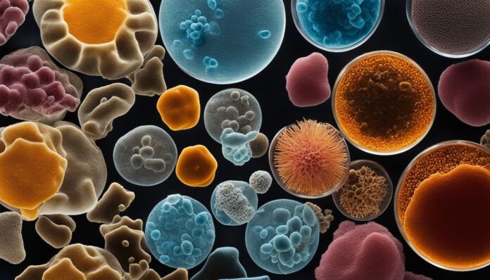
Bacteria are everywhere, tiny living things. They are hugely important in our health, making food, and our surroundings. Knowing techniques for the study of bacteria is key for scientists, doctors, and those in the microbiology field. In this detailed guide, you’ll learn about critical methods and tools. These are used to look at, find, and understand these tiny creatures. We will discuss everything from growing them to finding out who the bad ones are.
Key Takeaways
- Bacteria are everywhere, found in all kinds of life situations.
- Learning basic microbiology techniques is very important for experts in the field.
- The guide includes important steps for studying bacterial growth, counting, and recognizing them.
- We’ll talk about many topics, like how to handle bacteria safely and spot the dangerous ones.
- Getting good at these skills helps in making progress in areas such as healthcare, making new things, and taking care of our world.
Introduction to Microbiology Techniques
Bacteria live in many places and impact us in various ways. Some bacteria can make us very sick. Yet, others help us stay healthy and keep nature in balance. Knowing how to study bacteria helps in fields like medicine and protecting the environment.
Importance of Studying Bacteria
Bacteria are everywhere and are key to life on Earth. Learning how to see and understand them is crucial. It helps scientists, doctors, and others in the microbiology field to do their work.
Applications of Microbiology Techniques
Microbiology lab techniques are used in different areas. They help diagnose and treat bacterial diseases. They are also used to make antibiotics and even clean up polluted sites.
In addition, these methods are vital for learning more about life sciences. Bacteria allow us to study genetics and how living things function in a basic way.
Culturing and Aseptic Techniques
In microbiological work, it’s key to keep everything clean. We must protect tools, containers, and cultures from germs in the air. Aseptic technique makes sure everything is germ-free.
Aseptic Technique
To grow bacteria and fungi safely, you have to get things ready right. Clean the work area, wear the right gear, and set up what you need. This includes tools like an inoculating loop, stock cultures, and clean media.
Equipment and Work Area Preparation
Before making media, gather what you need. This could be a balance, spatula, and culture tubes. You might use dehydrated media or mix your own. It all needs to be sterilized, usually in an autoclave.
Media Preparation
You might mix your own media or use dehydrated kind. No matter what, you must sterilize it well. This kills any germs, making sure only the bacteria or fungi you want will grow.
Sterilization and Disinfection Methods
Keeping things sterile and clean is crucial in science labs and healthcare. There are three main ways to make sure things are safe to use: sterilizing, disinfecting, and sanitizing.
Sterilization Procedures
Some bacteria make spores that are really tough. They can survive high heat or boiling. To destroy these spores, we use steam under pressure. This high heat kills all harmful microorganisms. An autoclave is a common tool for this job.
Disinfection Techniques
Disinfection is killing or slowing the growth of germs on objects. We use chemicals or heat to do this. In labs, we may use things like chlorine or alcohol to clean. These substances differ in how well they fight different types of bacteria.
Sanitization Processes
Sanitization gets rid of harmful and not harmful germs on surfaces. It stops the spread of germs. Places like hospitals and restaurants use sanitization. It helps keep everyone safe and healthy by reducing the spread of diseases.
Isolation and Inoculation Techniques
In the world of microbiology, isolation and inoculation are key. They are vital for diagnostic tests and advanced research. These basics help researchers look at the bacteria around us.
Inoculation Methods
Inoculation is how we put a sample on a medium to grow cultures. Using a loop made of platinum or nichrome, we pick up a bit of a microbe. Putting this on the medium starts the growing process. Then, we can watch how the bacteria act.
Isolation of Microbial Strains
Isolation is essential for separating one strain from many in a culture. Placing them on specific growth mediums helps grow only the ones we want. This ensures we study the exact strain and exclude any other types. The goal is to have a pure culture for detailed study.
| Key Isolation and Inoculation Techniques | Description |
|---|---|
| Pour Plate Method | Adds a sample to agar to isolate and count microorganisms. This allows the count of colony-forming units (CFUs). |
| Spread Plate Method | Makes sure cells are spread for separate growth, aiding in enrichment and counting of microorganisms. |
| Streak Plate Method | Creates isolated colonies by streaking on a plate, allowing for clear colony growth. |
| Agar Stab Techniques | Uses a stab needle to make stab cultures for analyzing bacterial growth. |
Culture Media and Techniques
In the world of microbiology, growing bacteria is key. They grow in labs on culture media that have the right nutrients for them. Different types of media are used to grow specific microbes or to show their special traits.
Types of Culture Media
There are different types of culture media. These include simple, complex, and special media. Simple media have basic nutrients for many bacteria. For example, Tryptic Soy Agar (TSA) is a simple media. Complex media have extra nutrients, such as peptones and yeast extracts, for more picky microbes. These help the microbes grow better. Then we have synthetic or defined media. These have a known recipe and are usually found in research. They are very controlled. Special media are made for certain bacteria or fungi. Mannitol-Salt Agar (MSA) helps staphylococci bacteria grow, while Eosin-Methylene Blue Agar (EMB) shows which Gram-negative bacteria can ferment sugar.
Culture Techniques
Many techniques are used in microbiology labs. The streak plate, spread plate, and pour plate methods are common. They help scientists see one type of microbe at a time, count bacterial numbers, and grow pure cultures.
The streak plate method starts with spreading a sample on a culture medium. Then, you streak the sample to get isolated colonies. The spread plate method means putting a sample on the medium and spreading it out. The pour plate involves mixing a sample with melted agar, then pouring it into a dish to harden. This method captures microbes in the agar.
Techniques for the Study of Bacteria
Storing Microbial Samples
Researchers use several methods to store microbial samples for future study. These methods include refrigeration, deep freezing, lyophilization, and cryopreservation in liquid nitrogen. Each method helps maintain the health and qualities of the bacteria over time. This way, researchers can keep valuable samples for further analysis and research.
| Storage Technique | Description |
|---|---|
| Refrigeration | Storing microbial samples at low temperatures, typically between 2-8°C, to slow down metabolic processes and prevent growth. |
| Deep Freezing | Preserving microbial samples at ultra-low temperatures, usually below -70°C, to halt cellular activity and prevent degradation. |
| Lyophilization (Freeze-Drying) | Removing water from microbial samples through a process of freezing and desiccation, allowing for long-term storage at room temperature. |
| Cryopreservation in Liquid Nitrogen | Storing microbial samples in liquid nitrogen at -196°C, providing the most stable and long-term preservation solution for bacterial cultures. |
Bacterial Enumeration Methods
Finding the number of bacteria in a sample well is key in microbiology. It’s important for research and lab work. There are several ways to count these tiny organisms, and each method has its own use.
Serial Dilution
To make bacteria numbers manageable for study or culture, scientists dilute them. They dilute the sample in a series to find out how many microbes there are. They do this by counting the colonies that grow on plates.
Plate Counting Techniques
Scientists use methods like standard plate count and MPN to guess the number of live microbes. They put the diluted sample on special plates and count the colonies that form.
Spectrophotometric Analysis
With spectrophotometry, scientists can quickly measure the growth of microbes in a liquid. They measure how much light the bacteria absorb or scatter. This gives a precise count of the bacterial population.
| Enumeration Method | Principle | Key Features |
|---|---|---|
| Serial Dilution | Reducing bacterial concentration to countable levels | – Typically uses 10-fold or multiples of 10-fold dilutions – Ensures plates are not overgrown with colonies – Helps determine the number of microbial populations in a sample |
| Plate Counting Techniques | Inoculating culture media with diluted samples and counting colonies | – Standard plate count and most probable number (MPN) method – Provides information on viable bacterial cells – Colonies counted should ideally range from 30 to 300 for statistical accuracy |
| Spectrophotometric Analysis | Measuring the turbidity or optical density of a bacterial suspension | – Rapid and efficient method for estimating bacterial growth – Utilizes spectrophotometers or colorimeters to measure light absorption – Provides a quantitative assessment of bacterial populations |
These bacterial enumeration methods are crucial in the work of microbiology. They help experts precisely check the bacterial levels in many samples. This spans from food and water to clinical samples. Using these methods, researchers get vital info on bacterial communities, aiding progress in food science, environmental science, and biotechnology.
Identification of Bacterial Pathogens
Learning to spot bacterial pathogens is key to fighting infectious diseases. Scientists use lots of methods to tell different bacteria apart. Their main aim is to correctly diagnose and treat diseases caused by bacteria.
Morphological Examination
First, researchers look at how bacteria appear. They check their features like how they grow, their shape, and how big they are. This is done with the naked eye or a microscope. It’s a starting point for naming the type of bacteria based on its physical appearance.
Staining Techniques
Next, scientists might use stains to learn more. Different staining methods give clues about a bacteria’s cell wall composition and special parts inside. This helps to pin down the type of bacteria and even if it’s a dangerous pathogen.
For example, simple staining shows the basic shapes of bacteria. Gram staining looks at their cell walls to sort them into plus or minus groups. Then, there are special stains to pick out tough bacteria, like Mycobacterium. These cause serious diseases such as tuberculosis.
Combining visual checks and staining techniques is powerful. It helps scientists understand a lot about the bacteria they’re studying. This knowledge is crucial for deepening the identification and learning more about specific types of bacteria.
Advances in Bacterial Study Techniques
The world of microbiology is always changing. New methods and tools are making it easier to study bacteria. These steps include using genomic sequencing, microfluidics, and high-throughput screening.
These tools help us learn more about how bacteria work. They also show their genes and how they interact with the world around them. Microorganisms are a lot more than we thought.
Today, we’re able to look deeper into bacterial life than ever before. For example, advanced analysis methods like MALDI-TOF MS are making healthcare safer. We’re also using quick molecular tests to manage infectious diseases better.
Scientists are also testing new ways to understand microbe communities. This includes culturomics, metagenomic classification, and flow cytometry. As these new methods and improved “old” methods are used together, research in bacterial studies will keep advancing. This progress leads to better healthcare, safer public health efforts, and more effective environmental protection.
Source Links
- https://www.ncbi.nlm.nih.gov/pmc/articles/PMC8953368/
- https://conductscience.com/microbiology-techniques/
- https://www.ncbi.nlm.nih.gov/books/NBK8120/
- https://bio.libretexts.org/Courses/North_Carolina_State_University/MB352_General_Microbiology_Laboratory_2021_(Lee)/02:_Cultivation_of_Microbes/2.02:_Introduction_to_Bacterial_Growth_and_Aseptic_Techniques
- https://www.tmcc.edu/microbiology-resource-center/lab-protocols/aseptic-technique
- https://www.ncbi.nlm.nih.gov/pmc/articles/PMC7158362/
- https://www.colorado.edu/ehs/resources/disinfectants-sterilization-methods
- https://www.ncbi.nlm.nih.gov/pmc/articles/PMC7930362/
- https://www.tmcc.edu/microbiology-resource-center/lab-protocols/bacterial-isolation
- https://byjus.com/neet/inoculation/
- https://bio.libretexts.org/Learning_Objects/Laboratory_Experiments/Microbiology_Labs/Book:_Laboratory_Exercises_in_Microbiology_(McLaughlin_and_Petersen)/02:_Introduction_to_Aseptic_Techniques_and_Growth_Media/2.01:_Introduction
- https://conductscience.com/culture-media/
- https://www.ncbi.nlm.nih.gov/pmc/articles/PMC6961714/
- https://bio.libretexts.org/Courses/North_Carolina_State_University/MB352_General_Microbiology_Laboratory_2021_(Lee)/05:_Enumeration_of_Bacteria/5.01:_Introduction_to_Enumeration_of_Bacteria
- https://conductscience.com/bacteria-enumeration/
- https://study.com/academy/lesson/bacterial-enumeration-definition-methods-example.html
- https://www.ncbi.nlm.nih.gov/pmc/articles/PMC9769533/
- https://www.ncbi.nlm.nih.gov/pmc/articles/PMC3369584/
- https://www.ncbi.nlm.nih.gov/pmc/articles/PMC8207499/
- https://www.ncbi.nlm.nih.gov/pmc/articles/PMC6560418/


