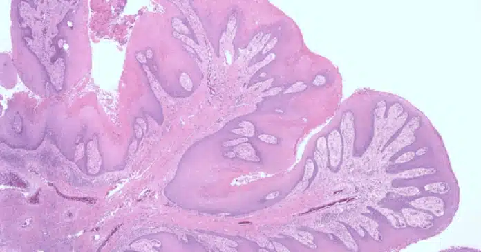
Histological staining is a cornerstone of cancer research and diagnosis, providing critical insights into the structure, composition, and behavior of cancer cells. By applying specific dyes or markers to tissue samples, researchers and pathologists can visualize cellular and molecular features that are otherwise invisible to the naked eye.
This process not only helps identify the presence of cancer but also reveals important details about tumor type, grade, and stage, guiding treatment decisions and improving patient outcomes.
In this blog post, we’ll explore the key histological staining techniques used in cancer research, from traditional methods like H&E staining to advanced immunohistochemistry and fluorescence staining, highlighting their role in cancer diagnosis, biomarker identification, and treatment development.
What is Histological Staining?
Histological staining involves the use of chemical dyes or antibodies to highlight specific components of tissue samples under a microscope. These stains interact with cellular structures, such as nuclei, cytoplasm, and extracellular matrix, to create contrast and reveal intricate details.
For example, Hematoxylin and Eosin (H&E) staining, the most widely used technique, colors nuclei blue-purple and cytoplasm pink, allowing pathologists to examine tissue architecture and detect abnormalities. In cancer research, staining techniques are tailored to identify malignant cells, assess tumor margins, and study the tumor microenvironment.
Importance of Staining in Cancer Diagnosis
Histological staining plays a pivotal role in cancer diagnosis by enabling the identification of cancerous cells and distinguishing them from healthy tissue. It helps pathologists determine the type of cancer (e.g., carcinoma, sarcoma, or lymphoma) and assess its aggressiveness.
For instance, Immunohistochemistry (IHC) uses antibodies to detect specific proteins, such as HER2 in breast cancer or PD-L1 in immunotherapy-responsive tumors. These insights are crucial for personalized treatment plans, as they reveal biomarkers that can predict how a patient will respond to therapy.
Overview of Common Staining Techniques
Several staining techniques are employed in cancer research, each with unique applications. H&E staining is the gold standard for initial tissue examination, providing a broad overview of tissue morphology. Special stains, such as Periodic Acid-Schiff (PAS) for carbohydrates or Masson’s trichrome for collagen, are used to highlight specific structures.
Advanced techniques like Fluorescence In Situ Hybridization (FISH) detect genetic abnormalities, while multiplex staining allows simultaneous visualization of multiple biomarkers. Together, these methods form a powerful toolkit for understanding cancer biology and improving diagnostic accuracy.
Key Histological Staining Techniques Used in Cancer Research
Histological staining techniques are the backbone of cancer research, enabling scientists and pathologists to visualize and analyze the complex structures of cancer cells and tissues. These techniques provide critical information about tumor type, grade, and molecular characteristics, which are essential for accurate diagnosis and effective treatment planning. Below, we explore some of the most widely used staining methods in cancer research and their unique applications.
Hematoxylin and Eosin (H&E) Staining
H&E staining is the most fundamental and widely used technique in histology. It involves two dyes: hematoxylin, which stains cell nuclei blue-purple, and eosin, which colors the cytoplasm and extracellular matrix pink. This simple yet powerful method provides a clear overview of tissue architecture, allowing pathologists to identify abnormal cell growth, tumor margins, and tissue damage. In cancer research, H&E staining is often the first step in diagnosing malignancies and determining the need for further specialized testing.
Immunohistochemistry (IHC) for Biomarker Detection
Immunohistochemistry (IHC) is a sophisticated staining technique that uses antibodies to detect specific proteins in tissue samples. These proteins, known as biomarkers, play a crucial role in cancer diagnosis and treatment. For example:
- HER2/neu is a biomarker for aggressive breast cancer, guiding the use of targeted therapies like trastuzumab.
- PD-L1 expression helps identify patients who may benefit from immunotherapy.
- Ki-67 is a proliferation marker used to assess tumor aggressiveness.
IHC provides valuable insights into the molecular profile of cancer cells, enabling personalized treatment strategies and improving patient outcomes.
Special Stains for Specific Cancer Types
In addition to H&E and IHC, special stains are used to highlight specific cellular components or structures that are relevant to particular types of cancer. Some commonly used special stains include:
- Periodic Acid-Schiff (PAS): Used to detect carbohydrates, PAS staining is helpful in diagnosing cancers like renal cell carcinoma and certain types of lymphoma.
- Masson’s Trichrome: This stain highlights collagen fibers, making it useful for studying the stroma of tumors, such as in pancreatic or liver cancer.
- Congo Red: Used to identify amyloid deposits, Congo red staining is essential in diagnosing amyloidomas, a rare type of tumor.
- Giemsa Stain: Often used in hematopathology, Giemsa staining helps differentiate blood cell types and diagnose hematologic malignancies like leukemia.
These specialized stains provide additional layers of information, enhancing the accuracy of cancer diagnosis and research.
Fluorescence In Situ Hybridization (FISH)
Fluorescence In Situ Hybridization (FISH) is a molecular staining technique that uses fluorescent probes to detect specific DNA sequences in cancer cells. FISH is particularly valuable for identifying genetic abnormalities, such as:
- Gene amplifications (e.g., HER2 in breast cancer).
- Chromosomal translocations (e.g., BCR-ABL in chronic myeloid leukemia).
- Deletions or duplications (e.g., EGFR mutations in lung cancer).
FISH provides high-resolution images of genetic alterations, making it a powerful tool for understanding the molecular basis of cancer and guiding targeted therapie
Applications of Histological Staining in Cancer Research
Histological staining is a vital tool in cancer research, providing researchers and clinicians with the ability to visualize and analyze tissue samples at a microscopic level. These techniques are not only essential for diagnosing cancer but also play a crucial role in understanding tumor biology, guiding treatment decisions, and monitoring disease progression. Below, we explore the key applications of histological staining in cancer research.
Tumor Grading and Staging
One of the primary applications of histological staining is in tumor grading and staging, which are critical for determining the aggressiveness and spread of cancer.
- Grading: Staining techniques like Hematoxylin and Eosin (H&E) allow pathologists to examine the cellular morphology and differentiate between low-grade (less aggressive) and high-grade (more aggressive) tumors. For example, the Gleason score in prostate cancer relies heavily on H&E staining to assess tumor patterns.
- Staging: Special stains and immunohistochemistry (IHC) help identify the extent of tumor invasion into surrounding tissues and lymph nodes, which is essential for staging cancers like breast, lung, and colorectal cancer.
Accurate grading and staging are crucial for developing personalized treatment plans and predicting patient outcomes.
Identifying the Tumor Microenvironment
The tumor microenvironment (TME) plays a significant role in cancer progression and treatment response. Histological staining techniques are used to study the complex interactions between cancer cells and their surrounding stroma, including immune cells, blood vessels, and extracellular matrix.
- IHC can identify immune cell markers (e.g., CD8+ T cells, macrophages) to assess the immune response within the tumor.
- Masson’s Trichrome staining highlights collagen fibers, providing insights into the stromal composition and its role in tumor growth and metastasis.
- Multiplex staining allows simultaneous visualization of multiple components of the TME, offering a comprehensive understanding of its dynamics.
Monitoring Treatment Response
Histological staining is invaluable for monitoring the effectiveness of cancer treatments, such as chemotherapy, radiation, and immunotherapy.
- IHC can detect changes in biomarker expression (e.g., HER2, PD-L1) before and after treatment, helping clinicians assess whether a therapy is working.
- H&E staining reveals morphological changes in tumor cells, such as necrosis or shrinkage, indicating treatment response.
- Fluorescence In Situ Hybridization (FISH) can track genetic alterations in response to targeted therapies, such as the reduction of HER2 amplification in breast cancer patients treated with trastuzumab.
Early Detection and Prevention
Histological staining also plays a critical role in the early detection and prevention of cancer.
- H&E staining is used to identify precancerous lesions, such as dysplasia in cervical or colorectal tissues, allowing for early intervention.
- Special stains like Periodic Acid-Schiff (PAS) can detect early changes in cellular structures, such as glycogen accumulation in liver cancer.
- IHC helps identify biomarkers associated with early-stage cancers, such as p53 mutations in precancerous lesions.
Early detection through histological staining can significantly improve survival rates by enabling timely treatment before cancer progresses to advanced stages.
Research and Development of New Therapies
Histological staining is a cornerstone of cancer research and drug development.
- Researchers use staining techniques to study the mechanisms of cancer progression, such as angiogenesis, invasion, and metastasis.
- IHC and FISH are used to validate the efficacy of new drugs in preclinical studies by assessing their impact on tumor cells and biomarkers.
- Advanced techniques like multiplex staining and 3D histology provide detailed insights into tumor biology, paving the way for innovative therapies.
By enabling a deeper understanding of cancer biology, histological staining accelerates the development of novel treatments and improves patient care.
Conclusion
Histological staining techniques are indispensable in cancer research, with applications ranging from diagnosis and treatment monitoring to understanding tumor biology and developing new therapies. By providing detailed visual and molecular insights into cancer cells and their microenvironment, these techniques empower researchers and clinicians to make informed decisions that improve patient outcomes. As technology advances, histological staining will continue to play a pivotal role in the fight against cancer, driving progress toward more effective and personalized treatments.


