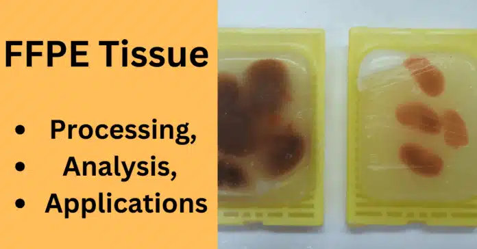
In the field of biomedical research and clinical diagnostics, FFPE tissue—short for Formalin-Fixed Paraffin-Embedded tissue—plays a crucial role. This widely used tissue preservation technique allows researchers and pathologists to store biological samples for long periods without compromising their structural integrity. By stabilizing tissues through formalin fixation and paraffin embedding, FFPE samples become ideal for detailed histopathological analysis, enabling the examination of cellular morphology and tissue architecture.
What makes FFPE tissue indispensable is its versatility. From cancer research and molecular profiling to biomarker discovery and retrospective studies using archival tissue samples, FFPE tissues provide a reliable resource for various applications. However, despite their advantages, FFPE samples present challenges, especially when isolating DNA, RNA, and proteins for molecular analyses due to potential nucleic acid degradation.
In this blog post, we will explore the entire journey of FFPE tissue—from its processing and analysis methods to its significant applications in oncology and personalized medicine. We will also discuss the key challenges researchers face when working with FFPE samples and highlight emerging solutions that are transforming how these tissues are utilized in modern science.
What is FFPE Tissue?
FFPE tissue, which stands for Formalin-Fixed Paraffin-Embedded tissue, is a standard method used in pathology and biomedical research for the long-term preservation of biological samples. This technique ensures that tissue morphology and cellular details remain intact for subsequent histological and molecular analyses. The process involves two main steps: formalin fixation, which stabilizes the tissue by cross-linking proteins, and paraffin embedding, which allows for easy slicing of the tissue into thin sections suitable for microscopic examination.
🔎 Why is FFPE Tissue Important?
FFPE tissues are considered the gold standard in histopathology because they preserve tissue architecture and cellular details crucial for disease diagnosis, especially in cancer research. Moreover, these tissues can be stored at room temperature for decades, making them invaluable for archival tissue repositories and retrospective studies. Researchers often rely on FFPE samples to study disease progression, identify biomarkers, and perform genomic and proteomic analyses using advanced techniques like next-generation sequencing (NGS) and PCR.
⚖️ FFPE vs. Fresh Frozen Tissue
When discussing tissue preservation techniques, it’s essential to compare FFPE tissue with fresh frozen tissue, another commonly used preservation method. While fresh frozen tissues better preserve nucleic acids and proteins for certain molecular analyses, they require strict storage conditions, such as ultra-low temperatures, which may not be feasible for long-term storage.
On the other hand, FFPE tissues are more practical for routine clinical diagnostics and histological studies, as they can be easily stored without specialized equipment. However, the formalin fixation process can lead to nucleic acid fragmentation and cross-linking, which pose challenges for molecular analysis. Recent advances in nucleic acid isolation protocols and antigen retrieval methods are helping overcome these challenges, making FFPE tissue an increasingly versatile resource.
🏥 Key Applications of FFPE Tissue
- Histological analysis for cancer diagnosis and classification.
- Molecular profiling to identify oncogenic pathways and genetic mutations.
- Immunohistochemistry (IHC) for detecting cancer biomarkers.
- Longitudinal studies using archival FFPE samples for retrospective research.
- Personalized medicine, where patient-specific FFPE samples guide therapeutic decisions.
FFPE Tissue Processing: Step-by-Step Guide
The processing of FFPE tissue is a critical procedure that ensures biological samples are preserved in a way that maintains their structural and molecular integrity for histological and molecular analyses. This process consists of several precise steps, including formalin fixation, paraffin embedding, tissue sectioning, and deparaffinization. Each step plays a crucial role in preparing the tissue for diagnostic and research applications, such as immunohistochemistry (IHC), DNA/RNA extraction, and protein analysis.
🧴 Step 1: Formalin Fixation
Formalin fixation is the first and most essential step in FFPE tissue processing. The tissue is immersed in a 10% neutral-buffered formalin solution, which acts as a fixative by cross-linking proteins and preserving cellular structures. This stabilization process prevents tissue degradation and maintains the sample’s morphology for histopathological analysis.
✅ Key Considerations for Optimal Fixation:
- Fixation Time: Typically 6–48 hours, depending on tissue size and type. Under-fixation can lead to tissue degradation, while over-fixation may cause excessive cross-linking, complicating nucleic acid isolation.
- Formalin Penetration: Ensuring uniform penetration is vital for consistent results in downstream applications like PCR and next-generation sequencing (NGS).
🕯️ Step 2: Paraffin Embedding
After fixation, the tissue undergoes paraffin embedding, which provides the structural support needed for microtome sectioning. The tissue is first dehydrated using increasing concentrations of alcohol to remove water, followed by treatment with xylene, which clears the tissue and makes it compatible with paraffin wax.
🔥 Embedding Process:
- Dehydration: Gradual exposure to ethanol solutions (70% to 100%).
- Clearing: Immersion in xylene to replace ethanol.
- Paraffin Infiltration: The tissue is saturated with molten paraffin wax.
- Embedding: The tissue is placed in a mold with paraffin and allowed to solidify, forming a block ready for sectioning.
✂️ Step 3: Tissue Sectioning
Once embedded, the FFPE tissue block is trimmed and sliced into thin sections using a microtome. These sections are typically 4–5 micrometers thick and are mounted onto glass slides for microscopic examination or further molecular analyses.
🎯 Considerations for Effective Sectioning:
- Blade Sharpness: Essential for clean, consistent sections.
- Section Thickness: Thinner sections are ideal for histological staining and IHC, while thicker sections may be required for DNA/RNA extraction and protein analysis.
- Adhesion to Slides: Treated slides improve section adhesion, reducing sample loss during deparaffinization and staining.
🧹 Step 4: Deparaffinization and Rehydration
Before molecular analysis or histological staining, the paraffin must be removed from the tissue sections through deparaffinization. This process typically involves immersing the slides in xylene, followed by a series of ethanol washes to rehydrate the tissue.
🧬 Deparaffinization Protocol:
- Xylene Treatment: Multiple immersions in xylene to dissolve paraffin.
- Rehydration: Stepwise exposure to decreasing ethanol concentrations (100% to 70%) and finally distilled water.
- Ready for Analysis: The rehydrated tissue is now prepared for Hematoxylin and Eosin (H&E) staining, IHC, or nucleic acid isolation for PCR and NGS.
🔧 Step 5: Antigen Retrieval (For IHC Applications)
Formalin fixation can mask antigenic sites, making them inaccessible to antibodies during immunohistochemistry (IHC). Antigen retrieval methods, such as heat-induced epitope retrieval (HIER) or enzyme-induced epitope retrieval (EIER), are used to reverse these cross-links, enhancing antigen accessibility and improving IHC results.
⚡ Step 6: Quality Control and Storage
Once processed, FFPE tissue sections undergo quality control checks, such as H&E staining to ensure structural integrity. Nucleic acid quality (e.g., RNA integrity number (RIN)) is also assessed when molecular analysis is required. Finally, FFPE tissue blocks are stored at room temperature in archival tissue repositories, where they can remain stable for years.
Analysis of FFPE Tissue: Methods and Challenges
The analysis of FFPE tissue is a crucial step in both clinical diagnostics and biomedical research, enabling the study of tissue morphology, disease mechanisms, and molecular biomarkers. However, working with FFPE samples presents specific challenges due to the tissue’s fixation and embedding process, which can affect the quality of DNA, RNA, and protein extracted from the tissue. Despite these challenges, advancements in molecular techniques and extraction protocols have made it possible to obtain valuable information from FFPE tissues for a variety of applications.
🧬 Molecular Analysis of FFPE Tissue
One of the primary uses of FFPE tissue is to perform molecular analysis, including DNA, RNA, and protein extraction for applications such as genetic mutation detection, gene expression profiling, and biomarker identification. However, the formalin fixation process can lead to nucleic acid fragmentation, making extraction and analysis more complex.
🔑 DNA Analysis
Despite the potential for DNA fragmentation, FFPE tissue remains a valuable resource for genetic analysis. Techniques such as PCR, quantitative PCR (qPCR), and next-generation sequencing (NGS) have been optimized to overcome DNA degradation. DNA quality is often assessed by measuring the fragment size and purity before proceeding with analysis.
🧪 RNA Analysis
RNA extraction from FFPE samples can be particularly challenging due to the cross-linking of nucleic acids during formalin fixation, which can result in RNA degradation. However, advances in RNA extraction kits have significantly improved the recovery of intact mRNA and non-coding RNAs, such as microRNAs. The use of reverse transcription followed by quantitative PCR (RT-qPCR) or RNA sequencing enables researchers to assess gene expression and identify molecular signatures from FFPE tissue.
🔬 Protein Analysis
Protein extraction from FFPE tissue is another area that requires special consideration due to the cross-linking of proteins by formalin. Techniques such as antigen retrieval and the use of protein extraction buffers are employed to break the cross-links and release proteins for subsequent proteomic analysis. Western blotting, mass spectrometry, and immunohistochemistry (IHC) are common methods used to study proteins in FFPE samples.
⚠️ Challenges in FFPE Tissue Analysis
While FFPE tissue offers invaluable material for various molecular analyses, several challenges need to be addressed to ensure accurate and reliable results.
💔 Nucleic Acid Degradation
The formalin fixation process, while preserving tissue morphology, often leads to nucleic acid fragmentation and cross-linking, making it difficult to extract high-quality DNA, RNA, and proteins. This can affect the sensitivity and specificity of downstream techniques like NGS and PCR. Researchers must use optimized extraction protocols and antigen retrieval methods to minimize the impact of this degradation.
⏳ Limited Sample Quantity
FFPE samples are often limited in quantity, especially when dealing with archival tissue samples or small biopsy samples. This constraint can limit the number of analytical tests that can be performed, requiring researchers to prioritize specific assays or methods.
🔬 PCR Inhibition
In some cases, paraffin wax residues may inhibit PCR amplification or other molecular techniques. This can be mitigated by using paraffin removal protocols and applying optimized DNA/RNA extraction kits specifically designed for FFPE samples.
🧪 Cross-Linking Effects on Proteins
The formalin fixation process can lead to protein cross-linking, which may reduce the accessibility of antigens for detection in techniques like IHC or Western blotting. To overcome this, researchers use antigen retrieval techniques to reverse the cross-linking and enhance the detection of specific proteins.
💡 Innovative Solutions to Overcome Challenges
Despite these challenges, significant advancements in the analysis of FFPE tissue have improved the reliability of results. Key innovations include:
- Improved DNA, RNA, and protein extraction kits specifically designed for FFPE tissue.
- Optimized antigen retrieval methods to enhance the accessibility of proteins and improve IHC results.
- Advances in next-generation sequencing (NGS) that allow for more efficient analysis of fragmented nucleic acids.
- Hybridization-based assays for gene expression profiling in degraded RNA.
These advances have expanded the range of molecular analyses that can be performed on FFPE tissue, making it a valuable resource for oncology, biomarker discovery, and personalized medicine.
Applications of FFPE Tissue in Research and Medicine
FFPE tissue is a cornerstone in both research and medicine due to its ability to preserve tissue samples for long periods without compromising morphological structure. It has become an indispensable resource in a wide range of fields, particularly in oncology, genomics, immunology, and biomarker discovery. The ability to perform molecular analyses on archival FFPE tissues has revolutionized the study of disease mechanisms, patient prognosis, and the development of personalized treatments.
🧬 1. Cancer Research
In cancer research, FFPE tissue plays a crucial role in understanding the genetic, molecular, and cellular basis of cancer. By enabling the preservation of tissue samples from cancer patients, FFPE specimens allow researchers to study tumor biology over time, uncover genetic mutations, and identify potential biomarkers for early cancer detection.
🔍 Applications in Cancer Research:
- Genetic Mutation Analysis: FFPE tissues are widely used to investigate mutations in oncogenes and tumor suppressor genes, such as TP53, KRAS, and EGFR, which are critical in the development and progression of various cancers.
- Biomarker Discovery: Researchers use FFPE tissue samples to identify molecular biomarkers that can help in early cancer detection, prognosis, and monitoring treatment efficacy.
- Gene Expression Profiling: Techniques like RNA sequencing (RNA-Seq) and quantitative PCR (qPCR) are applied to FFPE samples to evaluate the expression levels of genes involved in cancer-related pathways.
🧬 2. Diagnostic Applications
FFPE tissue is a critical resource in clinical diagnostics, particularly in pathology. Through immunohistochemistry (IHC) and molecular profiling, FFPE specimens enable the diagnosis of a wide array of diseases, including cancer, infectious diseases, and neurodegenerative conditions.
🧪 Diagnostic Uses in Medicine:
- Cancer Diagnosis and Classification: IHC allows pathologists to examine tissue sections for the presence of specific tumor markers, aiding in cancer diagnosis, tumor classification, and grading.
- Molecular Diagnostics: NGS and PCR-based assays on FFPE tissues can identify genetic mutations, fusion genes, and gene rearrangements, helping clinicians make accurate diagnoses and predict disease progression.
- Infectious Disease Detection: FFPE tissue samples can also be used to identify viral or bacterial infections at the molecular level, improving the diagnosis and treatment of infectious diseases.
🧬 3. Personalized Medicine
With the rise of personalized medicine, FFPE tissue has become a vital tool in developing tailored therapies based on a patient’s unique genetic and molecular profile. By analyzing FFPE samples for specific mutations or biomarkers, clinicians can identify which therapies are most likely to be effective for individual patients, particularly in cancer treatment.
🧑⚕️ Personalized Treatment Approaches:
- Targeted Therapies: FFPE tissue is used to identify targetable mutations, such as those in EGFR, BRAF, or ALK, which can guide the use of targeted therapies like tyrosine kinase inhibitors or monoclonal antibodies.
- Companion Diagnostics: FFPE samples play an essential role in companion diagnostics, where specific tests based on FFPE tissue help determine the suitability of a particular drug for a patient based on the molecular characteristics of their cancer.
- Immunotherapy: FFPE tissue analysis is also used to identify patients who might benefit from immune checkpoint inhibitors or other forms of immunotherapy, based on the expression of PD-L1 or other immune-related markers.
🧬 4. Biomarker Discovery and Development
The ability to extract high-quality DNA, RNA, and proteins from FFPE tissue has made it a valuable resource for the discovery and validation of biomarkers used in disease diagnosis, prognosis, and therapy. FFPE tissue is particularly useful in retrospective studies, where archived tissue samples can be analyzed for potential biomarkers that predict disease outcomes or response to treatment.
🔬 Applications in Biomarker Discovery:
- Cancer Biomarkers: FFPE samples are extensively used to discover cancer biomarkers related to tumor progression, drug resistance, and metastasis, which can be used for early detection or to monitor therapy efficacy.
- Therapeutic Targets: Proteomic analyses of FFPE tissue can help identify novel therapeutic targets, allowing for the development of drugs that specifically target disease-related proteins.
- Clinical Trials: FFPE tissue is often used in clinical trials to identify biomarkers that predict how patients will respond to specific therapies, aiding in the design of more effective clinical trials.
Keywords: Biomarker discovery, cancer biomarkers, therapeutic targets, proteomics, clinical trials, drug resistance, metastasis.
🧬 5. Disease Mechanisms and Pathogenesis
In addition to diagnostic and therapeutic applications, FFPE tissue provides researchers with the opportunity to study the underlying mechanisms of disease. By analyzing tissue samples from patients at different disease stages, researchers can gain insights into how diseases like cancer, neurodegenerative diseases, and cardiovascular conditions develop and progress.
🔍 Understanding Disease Mechanisms:
- Cancer Pathogenesis: FFPE tissue is used to study the molecular pathways involved in tumorigenesis, such as DNA damage repair, apoptosis, and angiogenesis.
- Neurodegenerative Diseases: FFPE tissue is also used to study diseases like Alzheimer’s and Parkinson’s disease, helping researchers identify early biomarkers and potential therapeutic targets.
- Cardiovascular Diseases: FFPE tissue samples from patients with cardiovascular diseases are analyzed to understand the molecular mechanisms underlying atherosclerosis, heart failure, and other cardiovascular conditions.
🌍 6. Global Health and Epidemiological Studies
FFPE tissue plays an important role in large-scale epidemiological studies, particularly those related to global health. By analyzing archival FFPE samples, researchers can track disease trends, identify risk factors, and assess the impact of environmental factors on health outcomes.
🌎 Applications in Epidemiology:
- Population Studies: FFPE tissue samples from large population-based studies help identify genetic predispositions to diseases like cancer, diabetes, and cardiovascular conditions.
- Environmental Exposures: Researchers use FFPE tissue to study the long-term effects of environmental exposures, such as pollution or occupational hazards, on disease development.
- Retrospective Cohort Studies: FFPE samples allow researchers to perform retrospective studies to understand how diseases progress and what interventions may be most effective.
Conclusion
FFPE tissue remains a cornerstone of medical research and diagnostics, offering invaluable insights into disease mechanisms and treatment responses. While working with FFPE samples comes with certain challenges, such as nucleic acid degradation, paraffin inhibition, and protein cross-linking, advancements in technology and optimized protocols have made it easier to overcome these obstacles. By leveraging improved extraction techniques, paraffin removal methods, and antigen retrieval strategies, researchers can continue to extract meaningful data from FFPE tissues, ensuring their continued relevance in clinical and research applications.


