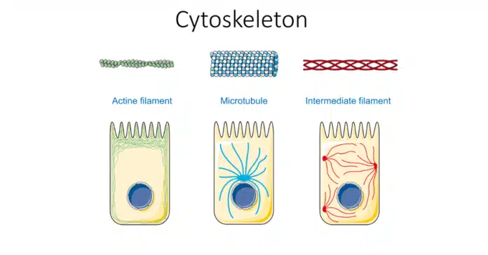
The cytoskeleton is a dynamic network of protein filaments crucial for maintaining cell shape, enabling movement, and ensuring mechanical support. It consists of microfilaments (actin filaments), microtubules, and intermediate filaments.
Unlike the more dynamic microfilaments and microtubules, intermediate filaments provide stable and resilient structural support. They are composed of proteins like keratins and vimentin, forming a robust network that helps cells withstand mechanical stress and maintain their integrity.
This article explores the various types and functions of intermediate filaments and compares them with other cytoskeletal components, including their role in plant cells.
What are Intermediate Filaments?
Intermediate filaments are one of the three main components of the cytoskeleton, alongside microfilaments and microtubules. They are named for their intermediate diameter, which is between the smaller microfilaments and the larger microtubules. Intermediate filaments typically measure about 10 nanometers in diameter, providing a balance between the flexibility of microfilaments and the rigidity of microtubules.
Definition and General Characteristics:
Intermediate filaments are strong, durable protein fibers that play a critical role in maintaining the structural integrity of cells and tissues.
They are less dynamic than microfilaments and microtubules, meaning they are more stable and not as frequently remodeled.
This stability allows them to provide long-term support and resilience to cells, especially those subjected to mechanical stress, such as skin cells and muscle cells.
Overall, intermediate filaments are essential for maintaining cellular architecture and integrity, particularly in cells that undergo significant mechanical strain. Their unique structural properties and diverse protein composition allow them to perform a variety of critical functions within different cell types.
Intermediate Filaments Examples
Intermediate filaments are diverse in their structure and function, tailored to meet the specific needs of different cell types. Here are the major types of them found in vertebrate cells, along with their component polypeptides and locations:
1. Nuclear Intermediate Filaments:
- Component Polypeptides: Lamins A, B, and C
- Location: These filaments form the nuclear lamina, which lines the inner surface of the nuclear envelope. Lamins provide structural support to the nucleus, maintaining its shape and stability. They are also involved in organizing chromatin and regulating DNA replication and cell division.
2. Vimentin-like Intermediate Filaments:
- Vimentin:
- Component Polypeptides: Vimentin
- Location: Found in many cells of mesenchymal origin, vimentin filaments help maintain cell shape, stabilize cytoplasmic organelles, and support cellular integrity. They play a crucial role in cell migration and wound healing.
- Desmin:
- Component Polypeptides: Desmin
- Location: Present in muscle cells, desmin filaments form a scaffold around the Z-disk in sarcomeres, providing structural integrity and aligning contractile elements. This ensures efficient force transmission during muscle contraction.
- Glial Fibrillary Acidic Protein (GFAP):
- Component Polypeptides: GFAP
- Location: Found in glial cells such as astrocytes and some Schwann cells, GFAP provides structural support and maintains the shape of these cells. It also plays a role in the functioning of the central nervous system.
- Peripherin:
- Component Polypeptides: Peripherin
- Location: Peripherin is found in some neurons, contributing to the structural integrity and function of the neuronal cytoskeleton.
3. Epithelial Intermediate Filaments:
- Type I Keratins (Acidic) and Type II Keratins (Neutral/Basic):
- Component Polypeptides: Type I keratins (acidic) and Type II keratins (neutral/basic)
- Location: Keratins are abundant in epithelial cells and their derivatives, such as hair and nails. They provide mechanical strength and protection against physical and chemical damage. Keratins form a robust network that helps epithelial tissues resist mechanical stress.
4. Axonal Intermediate Filaments:
- Neurofilament Proteins:
- Component Polypeptides: NF-L (light), NF-M (medium), and NF-H (heavy)
- Location: Found in neurons, neurofilaments are essential for maintaining the diameter of axons, which is crucial for the proper conduction of nerve impulses. They provide structural support to neurons and play a vital role in the development and maintenance of the nervous system.
Each type is uniquely adapted to its specific cellular context, providing mechanical support, maintaining cell shape, and ensuring proper cellular function.
This table summarizes the types of intermediate filaments, their component polypeptides, and their respective cellular locations.
| Type of Intermediate Filament | Component Polypeptides | Location |
|---|---|---|
| Nuclear Lamins | Lamins A, B, and C | Nuclear lamina (inner lining of nuclear envelope) |
| Vimentin-like | Vimentin | Many cells of mesenchymal origin |
| Desmin | Desmin | Muscle |
| Glial Fibrillary Acidic Protein | GFAP | Glial cells (astrocytes and some Schwann cells) |
| Peripherin | Peripherin | Some neurons |
| Epithelial Keratins | Type I keratins (acidic) and Type II keratins (neutral/basic) | Epithelial cells and their derivatives (e.g., hair and nails) |
| Axonal Neurofilament Proteins | NF-L, NF-M, and NF-H | Neurons |
Intermediate Filaments vs Actin Filaments (Microfilaments)
Structural Differences
Intermediate filaments and actin filaments (microfilaments) are both essential components of the cytoskeleton, but they differ significantly in their structure.
Intermediate filaments have a diameter of about 10 nanometers, which places them between the smaller actin filaments and the larger microtubules.
They are composed of elongated, fibrous proteins that form a coiled-coil dimer. These dimers associate in a staggered, antiparallel fashion to form tetramers, which then align end-to-end and laterally to create ropelike filaments.
In contrast, actin filaments are much thinner, with a diameter of about 7 nanometers. They are primarily composed of actin monomers, which polymerize to form long, thin, and flexible helical filaments, making them more dynamic than intermediate filaments.
Functional Roles in the Cell
Intermediate filaments provide structural stability and mechanical strength to cells. They form a durable network that helps cells withstand mechanical stress and maintain their shape.
This is especially important in cells that experience significant mechanical strain, such as epithelial cells, muscle cells, and neurons. They also play roles in positioning organelles within the cell and are involved in cellular processes like signal transduction and cell division.
Actin filaments (microfilaments), on the other hand, are involved in various dynamic cellular processes. They are essential for cell motility, enabling cells to move and change shape.
Actin filaments (microfilaments) are particularly crucial in muscle contraction, where they interact with myosin to produce force. They are also involved in cytokinesis, the process of cell division, where they form the contractile ring that separates the two daughter cells.
Additionally, actin filaments contribute to the formation of cellular extensions, such as filopodia and lamellipodia, which are important for cell migration and tissue development.
Comparison in Terms of Mechanical Properties and Cellular Processes
Intermediate filaments are more stable and less dynamic than actin filaments, providing long-term support and resilience to cells. They have high tensile strength, allowing them to endure mechanical stress without breaking.
This makes them well-suited for maintaining the structural integrity of cells and tissues under physical strain. Their primary role is to provide mechanical support and maintain cellular architecture.
In contrast, actin filaments are highly dynamic and constantly undergo polymerization and depolymerization. This dynamic nature allows them to quickly respond to changes in the cellular environment and participate in various cellular processes.
While actin filaments are not as strong as intermediate filaments, their flexibility and ability to rapidly assemble and disassemble make them ideal for functions that require quick and transient structural changes, such as cell movement, shape alterations, and muscle contraction.
Intermediate Filaments vs Microtubules
Structural Differences
Intermediate filaments and microtubules are two key components of the cytoskeleton, each with distinct structural characteristics. Microtubules are much larger, with a diameter of about 25 nanometers.
They are composed of tubulin dimers, which polymerize to form hollow tubes. Microtubules have a more rigid and tubular structure compared to the flexible, rope-like structure of intermediate filaments.
Functional Roles in the Cell
Microtubules are involved in various dynamic cellular processes. They are essential for maintaining cell shape, enabling intracellular transport, and facilitating cell division.
Microtubules form the mitotic spindle, which is crucial for chromosome segregation during mitosis. They also serve as tracks for the movement of organelles and vesicles within the cell, using motor proteins like kinesin and dynein to transport cargo.
Additionally, microtubules are involved in the formation of cilia and flagella, which are important for cell motility and sensory functions.
Comparison in Terms of Stability, Dynamics, and Cellular Processes
Microtubules are highly dynamic and constantly undergo polymerization and depolymerization. This dynamic behavior allows them to rapidly reorganize in response to cellular needs, making them ideal for functions that require quick structural changes.
Microtubules are less stable than intermediate filaments but are crucial for processes that require rapid assembly and disassembly, such as mitosis, intracellular transport, and the formation of cilia and flagella.
Intermediate Filaments in Plant Cells
Intermediate filaments are generally absent in plant cells, with the plant cytoskeleton being primarily composed of microtubules and actin filaments. Unlike animal cells, plants do not rely on intermediate filaments for structural support. However, plant cells have a unique structure called the plant cell wall, which provides mechanical strength and support, compensating for the lack of intermediate filaments.
Unique Functions in Plant Cellular Structure and Development: Although plant cells do not have intermediate filaments, they rely on the dynamic interactions between microtubules, actin filaments, and the cell wall to regulate cell shape, division, and expansion.
Microtubules, in particular, are involved in guiding the deposition of cellulose in the cell wall, influencing cell growth and development. Additionally, the plant cytoskeleton helps in organelle positioning and intracellular transport, ensuring proper cellular function and development.
Importance of Intermediate Filaments in Cellular Health
Intermediate filaments are essential for maintaining cell integrity by providing structural support and resilience against mechanical stress. They help cells retain their shape, protect against deformation, and ensure the proper positioning of organelles.
Involvement in Disease Conditions: Defects in intermediate filaments can lead to various diseases. For example, mutations in keratins can cause skin disorders like epidermolysis bullosa, while defects in neurofilaments are linked to neurodegenerative diseases such as amyotrophic lateral sclerosis (ALS). Additionally, disruptions in the function of intermediate filaments like desmin can result in muscle diseases, emphasizing their critical role in maintaining cellular health.
Conclusion
Intermediate filaments are essential components of the cytoskeleton, providing structural stability, mechanical support, and resilience to cells. They play crucial roles in maintaining cell integrity, supporting organelles, and facilitating cellular processes.
Unlike microfilaments and microtubules, intermediate filaments are more stable and less dynamic, but their disruption is linked to various diseases. Understanding their functions and significance highlights their importance in cellular health and disease prevention.


