

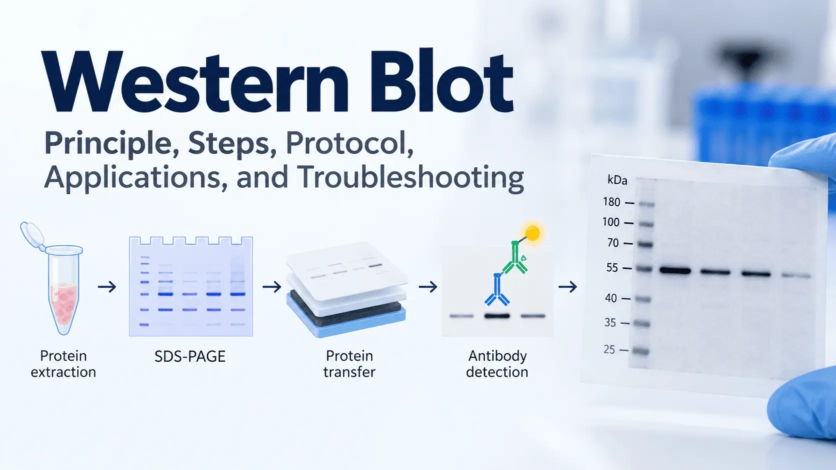
Western blot is one of the most widely used laboratory techniques for detecting and analyzing specific proteins in biological samples. It is commonly used in molecular biology, cell biology, immunology, biochemistry, and cancer research to determine whether a particular protein is present and to compare its relative abundance between samples.
The technique combines protein separation by electrophoresis with antibody-based detection. Proteins are first separated according to their molecular weight using SDS-PAGE, transferred to a membrane, and then detected using antibodies that specifically recognize the protein of interest.
Western blotting can provide valuable information about protein expression, signaling pathways, post-translational modifications, and changes in cellular processes. However, obtaining reliable results requires careful sample preparation, appropriate antibodies, efficient protein transfer, and suitable experimental controls.
This guide explains the Western blot principle, major steps, applications, data interpretation, and common troubleshooting strategies.
What Is Western Blot?
Western blot, also called Western blotting, is an analytical technique used to detect a specific protein within a complex mixture of proteins.
The technique relies on antibodies to recognize the target protein. Because thousands of proteins can be present in a biological sample, proteins are first separated according to their molecular weight by sodium dodecyl sulfate-polyacrylamide gel electrophoresis (SDS-PAGE). The separated proteins are then transferred from the gel onto a membrane, where they can be probed with specific antibodies.
The general workflow is:
Protein extraction → SDS-PAGE → Protein transfer → Blocking → Primary antibody → Secondary antibody → Detection → Data analysis
Western blotting is particularly useful when researchers want to determine whether a specific protein is expressed, compare protein abundance between experimental groups, or investigate changes in signaling pathways.
Western Blot vs. Southern and Northern Blot
The different blotting techniques are designed to analyze different types of biological molecules:
| Technique | Target molecule | Common application |
|---|---|---|
| Western blot | Protein | Protein detection and expression analysis |
| Southern blot | DNA | DNA sequence detection |
| Northern blot | RNA | RNA expression analysis |
Therefore, the defining feature of Western blotting is its ability to detect specific proteins using antibodies.
What Samples Can Be Used for Western Blot?
Western blotting can be performed using proteins extracted from a variety of biological samples, including:
- Cultured cells
- Animal tissues
- Human tissues
- Blood or biological fluids
- Cell lysates
- Tumor samples
- Subcellular fractions
The choice of sample depends on the biological question being investigated.
Western Blot Principle
The Western blot principle is based on separating proteins by electrophoresis, transferring them onto a membrane, and detecting a specific protein using antibodies.
The first step is to extract proteins from the biological sample. The protein mixture contains many different proteins with different molecular weights and concentrations.
The proteins are then mixed with a sample buffer containing SDS, which denatures proteins and gives them a relatively uniform negative charge. This allows their migration during electrophoresis to depend primarily on their molecular size.
Protein Separation by SDS-PAGE
During SDS-PAGE, the prepared protein samples are loaded into wells in a polyacrylamide gel.
When an electric field is applied, negatively charged proteins migrate through the gel toward the positive electrode. Smaller proteins generally move through the gel more rapidly than larger proteins.
As a result, proteins become separated according to their approximate molecular weight.
A protein molecular weight ladder is typically loaded alongside the samples to help estimate the size of the detected protein.
Transfer to a Membrane
After electrophoresis, the proteins are transferred from the polyacrylamide gel to a membrane.
Two commonly used membrane materials are:
- PVDF (polyvinylidene difluoride)
- Nitrocellulose
The transfer creates a stable protein-containing membrane that can be treated with antibodies.
Blocking
The membrane contains many sites where proteins or antibodies could bind nonspecifically. A blocking solution is therefore used to cover these nonspecific binding sites.
Common blocking reagents include nonfat milk and bovine serum albumin (BSA), although the appropriate blocker depends on the antibodies and experimental conditions.
Antibody-Based Detection
The membrane is incubated with a primary antibody that specifically recognizes the target protein.
A secondary antibody is then added. It recognizes the primary antibody and is usually linked to an enzyme or another detectable label.
Depending on the detection system, the signal can be visualized using chemiluminescence, fluorescence, or other detection methods.
The resulting band corresponds to the target protein.
Interpreting Protein Bands
The position of a Western blot band provides information about the approximate molecular weight of the detected protein.
The intensity of the band can provide information about the relative amount of protein between samples, provided that the experiment has been properly controlled and the signal is within an appropriate quantitative range.
Importantly, Western blot band intensity should not automatically be interpreted as an absolute measurement of protein concentration.
Western Blot Steps and Protocol
A typical Western blot protocol consists of several sequential steps. Conditions vary depending on the protein, antibodies, sample type, and laboratory system.
1. Protein Extraction
Proteins are extracted from cells or tissues using an appropriate lysis buffer.
The extraction procedure should preserve the target protein and minimize protein degradation. Protease inhibitors are commonly used when appropriate.
For proteins involved in signaling pathways, phosphatase inhibitors may also be required to preserve phosphorylation states.
2. Protein Quantification
Protein concentration is usually determined before loading the samples onto the gel.
Common protein assays include:
- Bradford assay
- BCA assay
- Other compatible colorimetric or fluorescence-based methods
Protein quantification helps ensure that comparable amounts of total protein are loaded between samples.
3. Sample Preparation and Denaturation
Protein samples are mixed with loading buffer containing SDS and reducing agents when appropriate.
Samples are commonly heated to promote protein denaturation.
The exact preparation conditions depend on the characteristics of the target protein because some proteins may require modified sample preparation.
4. SDS-PAGE
Prepared samples are loaded onto a polyacrylamide gel.
Electrophoresis separates proteins according to their molecular size. The appropriate gel percentage depends partly on the molecular weight range of the protein being studied.
A molecular weight marker should be included to help estimate protein size.
5. Protein Transfer
Following electrophoresis, proteins are transferred from the gel onto a PVDF or nitrocellulose membrane.
Transfer can be performed using different systems, including wet, semi-dry, and dry transfer approaches.
Transfer efficiency is an important factor because incomplete or uneven transfer can produce misleading results.
6. Membrane Blocking
The membrane is incubated with a blocking solution to reduce nonspecific antibody binding.
Blocking conditions should be optimized according to the antibody and detection system being used.
7. Primary Antibody Incubation
The membrane is incubated with a primary antibody directed against the target protein.
The antibody concentration and incubation conditions should follow the manufacturer’s recommendations as a starting point and can be optimized experimentally.
8. Washing
The membrane is washed to remove unbound or weakly associated primary antibody.
Proper washing is important for reducing background signal while maintaining specific antibody binding.
9. Secondary Antibody Incubation
A labeled secondary antibody that recognizes the primary antibody is applied to the membrane.
Secondary antibodies are commonly conjugated to enzymes such as horseradish peroxidase (HRP) or fluorescent labels.
10. Detection
After additional washing, the target protein is detected using an appropriate detection system.
Common approaches include:
- Chemiluminescence
- Fluorescence
- Colorimetric detection
The detected signal is captured using an imaging system.
11. Data Analysis
The resulting bands can be analyzed using densitometry.
The target protein signal is commonly normalized against an appropriate loading control or another validated normalization strategy.
The purpose is to determine whether protein abundance differs between experimental conditions.
Western Blot Applications in Research
Western blotting has numerous applications in biomedical and biological research.
Protein Expression Analysis
One of the most common applications is comparing protein expression between different samples or experimental conditions.
For example, researchers may compare the expression of a protein in treated and untreated cells.
Western blot can help determine whether an experimental treatment is associated with increased or decreased levels of a particular protein.
Analysis of Cell Signaling Pathways
Western blotting is widely used to investigate intracellular signaling pathways.
Researchers can measure proteins involved in pathways such as:
- PI3K/AKT
- MAPK/ERK
- JAK/STAT
- NF-κB
- mTOR
Phosphorylation-specific antibodies can also be used to investigate activation of signaling proteins.
For example, researchers may compare total AKT with phosphorylated AKT to investigate changes in AKT pathway activation.
Detection of Post-Translational Modifications
Western blotting can also be used to study post-translational modifications.
Depending on the available antibodies, researchers can investigate modifications such as:
- Phosphorylation
- Acetylation
- Ubiquitination
- Methylation
This makes Western blot particularly useful for studying changes in protein activity and regulation.
Western Blot in Cancer Research
Western blot is an important technique in cancer biology research.
Cancer cells frequently exhibit abnormal activation of signaling pathways that regulate proliferation, survival, metabolism, invasion, and migration.
Researchers can use Western blotting to investigate changes in proteins associated with these processes.
For example, Western blot may be used to examine:
- Oncogenic signaling pathways
- Tumor suppressor proteins
- Apoptosis-related proteins
- Cell-cycle regulators
- Epithelial-mesenchymal transition markers
- Angiogenesis-related proteins
- DNA damage response proteins
- Cancer stem cell markers
Western blotting can also be used to evaluate the molecular effects of potential anticancer treatments.
Validation of Gene Manipulation
Western blotting is often used to validate experiments involving gene overexpression or knockdown.
For example, after using siRNA to reduce expression of a particular gene, Western blot can determine whether the corresponding protein level has also decreased.
Similarly, Western blot can be used to evaluate whether gene overexpression results in increased protein production.
Biomarker Research
Western blot can contribute to biomarker research by evaluating candidate proteins in cells, tissues, or other biological samples.
However, Western blot results should be interpreted carefully, and additional experimental methods may be necessary to establish whether a protein is a clinically useful biomarker.
Western Blot Troubleshooting and Data Interpretation
Although Western blot is a powerful technique, researchers can encounter many technical problems.
Understanding common problems can help identify the source of poor-quality results.
No Bands
Possible causes include:
- Incorrect primary antibody
- Inactive antibody
- Incorrect antibody dilution
- Insufficient protein loading
- Poor protein transfer
- Incorrect detection conditions
- Degradation of the target protein
Checking each step systematically can help identify the problem.
Weak Bands
Weak signal may result from low target protein abundance, insufficient protein loading, excessive antibody dilution, inefficient transfer, or inadequate detection conditions.
Increasing the amount of loaded protein is not always the best solution because excessive protein loading can increase background and distort band intensity.
High Background
High background can make target bands difficult to distinguish.
Potential causes include:
- Inadequate blocking
- Excessive antibody concentration
- Insufficient washing
- Nonspecific antibody binding
- Incompatible blocking reagent
Optimization of antibody concentration and washing conditions can often improve the signal-to-background ratio.
Multiple or Nonspecific Bands
Multiple bands may occur when the antibody recognizes proteins other than the intended target.
Possible causes include antibody cross-reactivity, degradation products, alternative isoforms, or nonspecific binding.
Using a well-validated antibody and appropriate controls is particularly important when interpreting multiple bands.
Smearing
Protein smearing may be caused by:
- Protein degradation
- Overloading
- Poor sample preparation
- Problems during electrophoresis
- Inappropriate running conditions
- Poor-quality samples
Fresh protein samples and optimized electrophoresis conditions can help reduce this problem.
Uneven Protein Transfer
Uneven transfer can produce bands with inconsistent intensity across the membrane.
Potential causes include poor contact between the gel and membrane, air bubbles, inappropriate transfer conditions, or differences in gel and membrane preparation.
Careful assembly of the transfer sandwich is therefore important.
Loading Controls
Loading controls are commonly used to assess whether similar amounts of protein were loaded across samples.
Frequently used controls include:
- β-actin
- GAPDH
- α-tubulin
However, the choice of loading control should be experimentally justified because the expression of housekeeping proteins can change under certain experimental conditions.
Densitometric Analysis
Western blot data are often quantified by measuring the intensity of protein bands.
The target protein signal can be normalized to an appropriate loading control or other validated normalization method.
For example:
Normalized protein expression = Target protein signal / Loading control signal
The normalized values can then be compared between experimental groups.
It is important to avoid interpreting saturated bands quantitatively because signal saturation can distort differences in protein abundance.
Importance of Experimental Controls
Reliable Western blot experiments require appropriate controls.
Depending on the experimental design, these may include:
- Positive controls
- Negative controls
- Untreated controls
- Loading controls
- Molecular weight markers
- Secondary-antibody-only controls
Controls help determine whether an observed signal represents the target protein rather than a technical artifact.
Conclusion
Western blot is a fundamental technique for detecting and analyzing specific proteins in biological samples. Its principle combines SDS-PAGE-based protein separation with membrane transfer and antibody-mediated detection.
The main stages include protein extraction, protein quantification, SDS-PAGE, membrane transfer, blocking, primary and secondary antibody incubation, detection, and data analysis.
The technique has broad applications in molecular biology, cell biology, immunology, and cancer research. It can be used to investigate protein expression, signaling pathways, post-translational modifications, gene manipulation, and molecular mechanisms of disease.
However, obtaining reliable Western blot results requires careful experimental design. Antibody specificity, protein quality, transfer efficiency, appropriate controls, and proper data normalization are all essential for meaningful interpretation.
For researchers and students working in biomedical sciences, understanding the principle, protocol, applications, and troubleshooting of Western blotting provides an important foundation for studying protein biology and cellular mechanisms.


