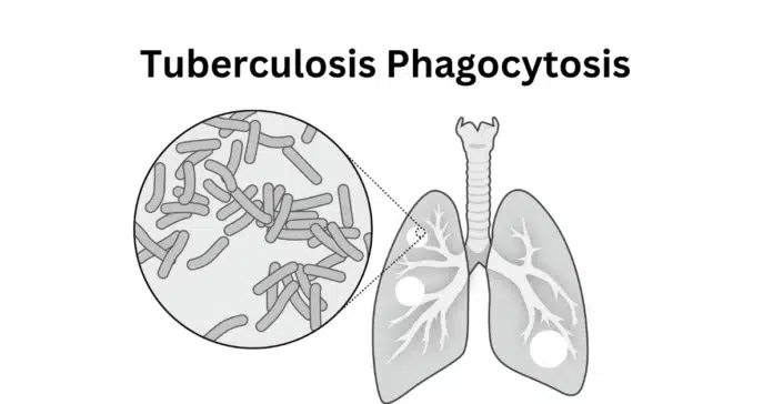
In this article, we will explore the mechanism of tuberculosis phagocytosis, how M. tuberculosis manipulates the immune system, and the potential therapeutic strategies aimed at overcoming its evasion tactics. Understanding these processes is essential for developing better treatments and vaccines to combat tuberculosis.
Tuberculosis (TB) remains one of the deadliest infectious diseases worldwide, caused by the bacterium Mycobacterium tuberculosis. This pathogen primarily infects the lungs but can spread to other organs, leading to severe complications.
A key factor in TB’s success as a human pathogen is its ability to manipulate the immune system—especially through interactions with macrophages, the first line of defense against infection.
Phagocytosis, the process by which immune cells engulf and destroy pathogens, is crucial in containing bacterial infections. However, M. tuberculosis has evolved sophisticated mechanisms to evade destruction within macrophages, allowing it to persist in the host and establish either active or latent tuberculosis.
By inhibiting phagosome maturation, resisting reactive oxygen species (ROS), and modulating the immune response, the bacterium creates a survival niche within host cells.
2. What is Phagocytosis in Tuberculosis?
Phagocytosis is a fundamental process of the innate immune system, where specialized immune cells, such as macrophages, dendritic cells, and neutrophils, engulf and eliminate pathogens. In the case of tuberculosis, phagocytosis is the first line of defense against Mycobacterium tuberculosis (M. tuberculosis), as alveolar macrophages in the lungs recognize and attempt to destroy the invading bacteria.
The Role of Macrophages in Tuberculosis Infection
Macrophages are the primary host cells for M. tuberculosis. Once the bacteria are inhaled and reach the alveoli, macrophages recognize them through pattern recognition receptors (PRRs) such as:
- Toll-like receptors (TLRs) – Detect bacterial components and trigger immune signaling.
- Mannose receptors (MRs) – Bind to M. tuberculosis surface mannose molecules.
- Complement receptors (CR3, CR4) – Aid in bacterial uptake via opsonization.
After recognition, the macrophage engulfs M. tuberculosis into a phagosome, an intracellular vesicle designed to fuse with lysosomes, forming a phagolysosome. In normal infections, this process results in pathogen degradation via:
- Acidification of the phagolysosome
- Release of reactive oxygen species (ROS) and reactive nitrogen species (RNS)
- Enzymatic breakdown of bacterial components
However, M. tuberculosis has evolved strategies to disrupt this process, preventing its destruction and allowing intracellular survival. This immune evasion mechanism is what makes tuberculosis particularly challenging to treat.
3. Mechanism of Mycobacterium tuberculosis Phagocytosis
Phagocytosis is a multi-step process involving the recognition, engulfment, and digestion of pathogens. However, when it comes to Mycobacterium tuberculosis (M. tuberculosis), this process is not entirely straightforward. While macrophages efficiently recognize and engulf the bacteria, M. tuberculosis has developed sophisticated mechanisms to manipulate and evade immune destruction inside the host cell.
Step 1: Recognition and Binding of M. tuberculosis
Upon inhalation, M. tuberculosis reaches the alveoli, where it is recognized by alveolar macrophages. The bacteria interact with various host cell surface receptors, which facilitate their uptake:
- Toll-like receptors (TLRs) – Activate pro-inflammatory responses upon bacterial detection (e.g., TLR2, TLR4, and TLR9).
- Mannose receptors (MRs) – Recognize mannosylated molecules on M. tuberculosis surface, aiding its internalization.
- Complement receptors (CR3, CR4) – Enable bacterial entry via complement-opsonized pathways.
- Fc receptors (FcRs) – Bind to antibody-coated bacteria and mediate phagocytosis.
Despite these immune mechanisms, M. tuberculosis actively modulates receptor signaling to minimize macrophage activation, reducing the effectiveness of the immune response.
Step 2: Engulfment and Phagosome Formation
Once recognized, M. tuberculosis is engulfed into a membrane-bound vesicle known as a phagosome. This step is energy-dependent and involves:
- Actin polymerization for cytoskeletal rearrangement.
- Formation of a phagocytic cup that gradually closes around the bacterium.
- Internalization of the pathogen into the early phagosome, which is supposed to mature into a bactericidal phagolysosome.
However, M. tuberculosis has evolved mechanisms to arrest this process, allowing its survival inside macrophages.
Step 3: Phagosome-Lysosome Maturation Arrest
Under normal conditions, phagosomes fuse with lysosomes to form phagolysosomes, where bacteria are degraded through:
- Acidification (via V-ATPase proton pumps).
- Hydrolytic enzyme activity.
- Production of reactive oxygen species (ROS) and reactive nitrogen species (RNS).
However, M. tuberculosis blocks this maturation process, preventing lysosomal fusion and avoiding destruction. The bacterium does this by:
Interfering with Phagosome Acidification
- M. tuberculosis secretes LAM (lipoarabinomannan), which inhibits V-ATPase recruitment, preventing acidification.
Blocking Phagosome-Lysosome Fusion
- The bacterium modifies phagosomal membrane proteins, preventing lysosomal enzyme delivery.
- It secretes PtpA, a phosphatase that disrupts host signaling, halting lysosomal fusion.
Escaping Reactive Oxygen and Nitrogen Species (ROS & RNS)
- M. tuberculosis produces Superoxide Dismutase (SOD) and Catalase-Peroxidase (KatG) to neutralize oxidative stress.
- It also secretes NuoG, which inhibits apoptosis, allowing prolonged intracellular survival.
Step 4: Survival and Intracellular Persistence
By inhibiting normal macrophage functions, M. tuberculosis establishes a persistent infection. Some key survival strategies include:
- Inhibition of apoptosis: M. tuberculosis prevents programmed cell death, keeping its host cell alive longer.
- Manipulation of cytokine production: The bacterium suppresses pro-inflammatory signals (TNF-α, IFN-γ) while enhancing anti-inflammatory cytokines (IL-10) to avoid immune detection.
- Formation of granulomas: Infected macrophages form granulomas, which help contain the infection but also provide a protective niche for M. tuberculosis.
In the next section, we will explore how M. tuberculosis further evades immune destruction and survives within host cells.
4. How Mycobacterium tuberculosis Evades Phagocytosis
Mycobacterium tuberculosis (M. tuberculosis) has evolved highly specialized mechanisms to evade destruction within macrophages, allowing it to persist inside the host and establish either latent or active tuberculosis. Unlike most pathogens that are destroyed upon phagocytosis, M. tuberculosis actively modifies host cell processes to prevent its elimination.
Below are the key immune evasion strategies employed by M. tuberculosis:
1. Inhibition of Phagosome-Lysosome Fusion
Normally, after a pathogen is engulfed, the phagosome matures and fuses with lysosomes, forming a phagolysosome where the bacteria are destroyed by hydrolytic enzymes and acidic pH. However, M. tuberculosis blocks this process by:
Secreting Lipoarabinomannan (LAM):
- LAM, a component of the bacterial cell wall, inhibits the recruitment of V-ATPase, preventing phagosome acidification.
- This results in a neutral pH environment, allowing the bacterium to survive.
Blocking SNARE Proteins Involved in Fusion:
- The bacterium modifies phagosomal membrane proteins, preventing fusion with lysosomes.
- It secretes PtpA (Protein Tyrosine Phosphatase A), which disrupts host signaling pathways necessary for lysosome recruitment.
Interfering with Host GTPases:
- M. tuberculosis alters the function of Rab GTPases, which control vesicle trafficking, further preventing lysosome fusion.
2. Resistance to Reactive Oxygen and Nitrogen Species (ROS & RNS)
Macrophages produce reactive oxygen species (ROS) and reactive nitrogen species (RNS) as antimicrobial defenses to kill engulfed pathogens. M. tuberculosis, however, has evolved powerful antioxidant mechanisms to neutralize these toxic molecules:
Production of Superoxide Dismutase (SOD) and Catalase-Peroxidase (KatG):
- These enzymes detoxify ROS, preventing oxidative damage to the bacterium.
Secretion of NuoG:
- NuoG blocks NO-induced apoptosis, allowing M. tuberculosis to persist inside the macrophage.
Downregulation of iNOS (Inducible Nitric Oxide Synthase):
- M. tuberculosis actively suppresses iNOS expression, reducing nitric oxide (NO) production, a key antimicrobial defense.
3. Manipulation of the Host Immune Response
To avoid detection and destruction by the immune system, M. tuberculosis actively modulates cytokine signaling and host immune responses:
Inhibition of Pro-Inflammatory Cytokines (TNF-α, IFN-γ):
- M. tuberculosis suppresses TNF-α and IFN-γ, two critical cytokines involved in macrophage activation and bacterial clearance.
Induction of Anti-Inflammatory Cytokines (IL-10, TGF-β):
- The bacterium promotes IL-10 and TGF-β production, which dampen immune responses and create a permissive intracellular environment.
Interference with Antigen Presentation:
- M. tuberculosis reduces MHC-II expression, preventing effective T-cell activation and adaptive immune response.
4. Prevention of Macrophage Apoptosis and Induction of Necrosis
Apoptosis is a host defense mechanism that eliminates infected cells while limiting bacterial spread. However, M. tuberculosis actively suppresses apoptosis and promotes necrosis, which helps it escape the host cell and infect neighboring cells.
Inhibition of Apoptosis:
- M. tuberculosis blocks apoptotic pathways by upregulating anti-apoptotic proteins (Bcl-2, Mcl-1) and downregulating pro-apoptotic signals.
Induction of Necrosis:
- Instead of undergoing programmed cell death, infected macrophages rupture, releasing bacteria into surrounding tissues and promoting further infection.
5. Formation of Granulomas: A Double-Edged Sword
As a defense mechanism, the immune system forms granulomas—organized immune cell structures that contain the bacteria. While granulomas initially restrict bacterial spread, M. tuberculosis exploits them as a survival niche by:
Surviving in Dormant Form:
- The bacterium can enter a non-replicative state, allowing long-term persistence (latent TB).
Reactivating When Immunity Weakens:
- If the immune system becomes compromised (e.g., HIV infection, aging), M. tuberculosis reactivates, leading to active TB disease.
5. Role of Phagocytosis in Tuberculosis Granuloma Formation
Granulomas are a hallmark of tuberculosis (TB) and play a dual role in infection: they help contain Mycobacterium tuberculosis (M. tuberculosis) but also provide a niche for bacterial persistence. Phagocytosis plays a crucial role in granuloma formation, as macrophages and other immune cells interact to control the infection.
1. What is a Tuberculosis Granuloma?
A granuloma is a structured aggregation of immune cells formed in response to persistent infections like TB. It consists of:
- Infected macrophages (which phagocytose M. tuberculosis)
- Foamy macrophages (lipid-laden cells that support bacterial survival)
- Multinucleated giant cells (fused macrophages)
- Epithelioid cells (differentiated macrophages with barrier functions)
- T cells (which regulate macrophage activity)
- Fibroblasts (which contribute to tissue remodeling)
2. Phagocytosis as the Initiating Event in Granuloma Formation
The process begins when alveolar macrophages recognize and phagocytose M. tuberculosis after inhalation. This leads to:
Macrophage Activation:
- Phagocytosis triggers pro-inflammatory signals (TNF-α, IFN-γ), recruiting more immune cells.
- Activated macrophages attempt to destroy the bacteria but often fail due to M. tuberculosis‘s evasion strategies.
Recruitment of Immune Cells:
- Dendritic cells and macrophages present antigens to T cells, promoting immune activation.
- Neutrophils and monocytes are attracted to the site, enhancing phagocytosis.
Formation of the Granuloma Core:
- Infected macrophages cluster together, forming the central core of the granuloma.
- Some macrophages differentiate into foamy macrophages, which store lipids and provide a nutrient-rich environment for M. tuberculosis.
Giant Cell and Epithelioid Cell Formation:
- Macrophages fuse into multinucleated giant cells to restrict bacterial spread.
- Some macrophages transform into epithelioid cells, creating a physical barrier.
3. How Granulomas Contain M. tuberculosis
Initially, granulomas function as a protective immune barrier:
Restricting bacterial spread: The physical structure of the granuloma traps bacteria, preventing systemic dissemination.
Inducing immune activation: Granulomas contain activated macrophages that produce reactive oxygen species (ROS) and nitric oxide (NO) to kill bacteria.
Promoting adaptive immunity: T cells within granulomas secrete IFN-γ to stimulate macrophages and enhance bacterial clearance.
However, M. tuberculosis can exploit granulomas for survival instead of being eliminated.
4. How M. tuberculosis Exploits Granulomas for Persistence
Despite their protective role, granulomas can become a reservoir for M. tuberculosis due to:
Failure of Phagosome-Lysosome Fusion:
- Macrophages inside granulomas fail to fully kill M. tuberculosis, allowing bacterial survival.
Induction of Foamy Macrophages:
- M. tuberculosis manipulates lipid metabolism, creating foamy macrophages that provide a nutrient-rich niche for bacterial persistence.
Granuloma Necrosis and Bacterial Escape:
- Over time, some granulomas undergo necrosis, breaking down and releasing live M. tuberculosis into the lungs.
- This leads to cavitary TB, where bacteria are expelled in aerosols and transmitted to new hosts.
5. Granulomas in Latent vs. Active TB
Granulomas play a key role in determining disease progression:
Latent TB (Controlled Granulomas):
- M. tuberculosis is contained within granulomas in a dormant state.
- A strong immune response prevents bacterial escape, keeping the infection asymptomatic.
Active TB (Breakdown of Granulomas):
- In immunocompromised individuals, granulomas break down, releasing bacteria into the airways.
- This leads to chronic cough, lung tissue destruction, and contagious TB disease.
6. Potential Therapeutic Strategies Targeting Tuberculosis Phagocytosis
Since Mycobacterium tuberculosis (M. tuberculosis) can evade phagocytosis and persist within macrophages, targeting the phagocytic process presents a promising therapeutic strategy. Current research focuses on enhancing macrophage function, restoring phagosome-lysosome fusion, and boosting host immune responses to eliminate M. tuberculosis.
1. Enhancing Tuberculosis Phagocytosis and Macrophage Activation
Strengthening macrophage activity can improve bacterial clearance and prevent TB progression.
Immunotherapy with IFN-γ (Interferon-Gamma)
- IFN-γ enhances macrophage activation, promoting phagocytosis and intracellular bacterial killing.
- Used as an adjunct therapy in patients with multidrug-resistant TB (MDR-TB).
Boosting Toll-Like Receptor (TLR) Signaling
- TLRs recognize M. tuberculosis and trigger macrophage activation.
- Synthetic TLR agonists (e.g., CpG ODNs, Pam3CSK4) enhance phagocytosis and cytokine production.
Autophagy-Inducing Compounds
- Rapamycin and Metformin stimulate autophagy, a process that degrades intracellular bacteria.
- Autophagy-enhancing drugs restore phagosome-lysosome fusion, improving bacterial clearance.
2. Restoring Phagosome-Lysosome Fusion
One of M. tuberculosis’s key evasion strategies is blocking phagosome-lysosome fusion. New approaches aim to counteract this:
Targeting PtpA (Protein Tyrosine Phosphatase A)
- M. tuberculosis secretes PtpA, which prevents lysosome recruitment.
- PtpA inhibitors restore normal phagosomal maturation, allowing bacterial degradation.
Stimulating V-ATPase Activity
- The V-ATPase complex acidifies lysosomes, promoting bacterial destruction.
- Drugs that boost V-ATPase activity enhance lysosomal function in macrophages.
Host-Directed Therapy with Vitamin D
- Vitamin D promotes cathelicidin (LL-37) production, which enhances phagosome maturation and bacterial killing.
- Used in clinical trials as an adjunct therapy for TB patients.
3. Modulating the Inflammatory Response
Balancing the immune response is crucial—excessive inflammation can cause tissue damage, while insufficient activation allows bacterial persistence.
TNF-α Modulation
- TNF-α is essential for granuloma formation and macrophage activation, but excessive levels cause tissue damage.
- Selective TNF-α modulators (not complete inhibitors) may help maintain an effective immune response.
Blocking IL-10 to Restore Macrophage Function
- M. tuberculosis upregulates IL-10, an anti-inflammatory cytokine that suppresses macrophage activity.
- IL-10 inhibitors may enhance phagocytosis and intracellular bacterial clearance.
4. Host-Directed Therapies (HDTs) for Tuberculosis Phagocytosis
Host-directed therapies (HDTs) target the host’s immune response instead of directly attacking M. tuberculosis.
Metformin (Diabetes Drug) as an Immune Booster
- Metformin enhances AMPK (AMP-activated protein kinase) signaling, which boosts autophagy and bacterial clearance.
- Clinical studies suggest metformin improves TB treatment outcomes.
Statins to Enhance Macrophage Function
- Statins have anti-inflammatory and autophagy-enhancing effects.
- They promote phagosome-lysosome fusion and improve TB treatment responses.
Nutritional Supplements (Vitamin D & Omega-3 Fatty Acids)
- Vitamin D enhances macrophage antimicrobial activity.
- Omega-3 fatty acids modulate inflammation and improve phagocytosis.
5. Nanotechnology-Based Drug Delivery for Macrophage Targeting
Since M. tuberculosis hides inside macrophages, delivering drugs directly to infected cells can improve treatment efficacy.
Nanoparticle-Encapsulated Anti-TB Drugs
- Lipid-based nanoparticles (liposomes) can target infected macrophages more efficiently than standard TB drugs.
- PLGA nanoparticles (polymeric nanoparticles) improve drug stability and delivery to granulomas.
Host-Targeted siRNA Therapy
- RNA-based therapies silence host genes that M. tuberculosis exploits for survival (e.g., Rab GTPases, PtpA).
- Experimental studies show siRNA restores normal phagosome function.
Targeting TB phagocytosis and macrophage function offers promising new therapeutic strategies. While traditional antibiotics focus on killing bacteria, host-directed therapies aim to boost immune responses and restore phagosome maturation.
Current Research Focus:
Developing drugs that enhance phagocytosis
Targeting M. tuberculosis’s evasion strategies
Using host-directed therapies to improve TB treatment outcomes
Conclusion
Phagocytosis plays a crucial role in tuberculosis, serving as both a defense mechanism and a target for Mycobacterium tuberculosis evasion strategies. While macrophages attempt to eliminate the bacteria, M. tuberculosis manipulates this process to survive and persist within granulomas. Understanding these interactions has paved the way for innovative therapeutic strategies, including host-directed therapies, immunomodulators, and nanotechnology-based drug delivery systems. Targeting phagocytosis could enhance TB treatment outcomes, offering new hope in the fight against this global health threat.
FAQ: Tuberculosis Phagocytosis
1. How does TB survive phagocytosis?
Mycobacterium tuberculosis (M. tuberculosis) has evolved multiple mechanisms to survive inside macrophages after being engulfed through phagocytosis. It prevents phagosome-lysosome fusion, neutralizes reactive oxygen species (ROS), and exploits the host’s lipid metabolism to create a nutrient-rich environment within macrophages. These strategies allow the bacteria to persist instead of being destroyed.
2. Why is phagocytosis not always effective in fighting TB?
Phagocytosis alone is often insufficient to eliminate M. tuberculosis because the bacteria can manipulate immune signaling pathways. It blocks key antimicrobial responses, suppresses macrophage activation, and creates a persistent infection inside granulomas. In some cases, an excessive immune response leads to tissue damage instead of bacterial clearance.
3. How does TB prevent phagolysosome fusion?
M. tuberculosis secretes specific proteins, such as PtpA and SapM, which interfere with phagosome maturation and prevent lysosomal fusion. This allows the bacterium to remain in a safe, non-acidic environment, where it can replicate and avoid degradation by macrophage enzymes.
4. How does tuberculosis invade the immune system?
M. tuberculosis invades the immune system by targeting alveolar macrophages in the lungs. It disrupts normal immune responses by:
Inhibiting antigen presentation to avoid detection by T cells.
Inducing IL-10 production, which suppresses macrophage activity.
Modulating granuloma formation, creating a niche for bacterial persistence.
Escaping into the extracellular space when granulomas break down, leading to active disease and transmission.


